

DEC-205-mediated antigen targeting to steady-state dendritic cells induces deletion of diabetogenic CD8⁺ T cells independently of PD-1 and PD-L1
- PMID: 24021877
- PMCID: PMC3806169
- DOI: 10.1093/intimm/dxt031
DEC-205-mediated antigen targeting to steady-state dendritic cells induces deletion of diabetogenic CD8⁺ T cells independently of PD-1 and PD-L1
Abstract
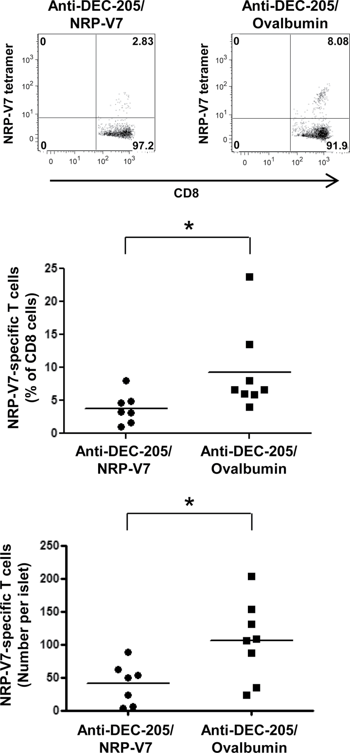
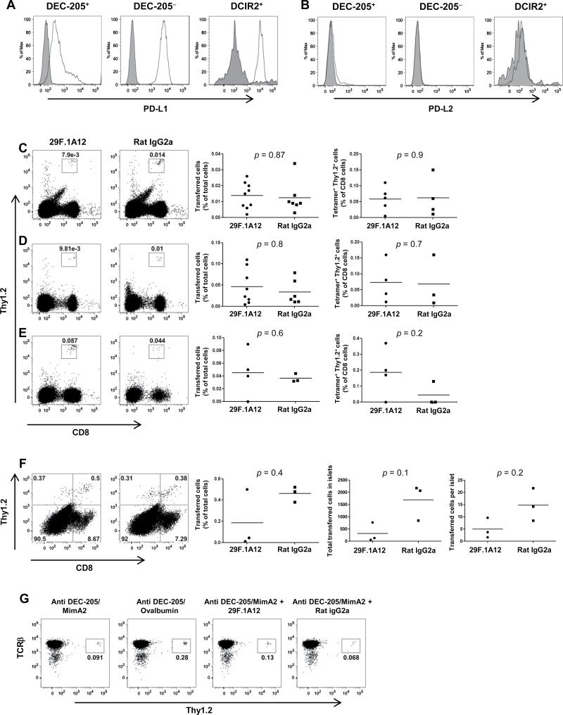
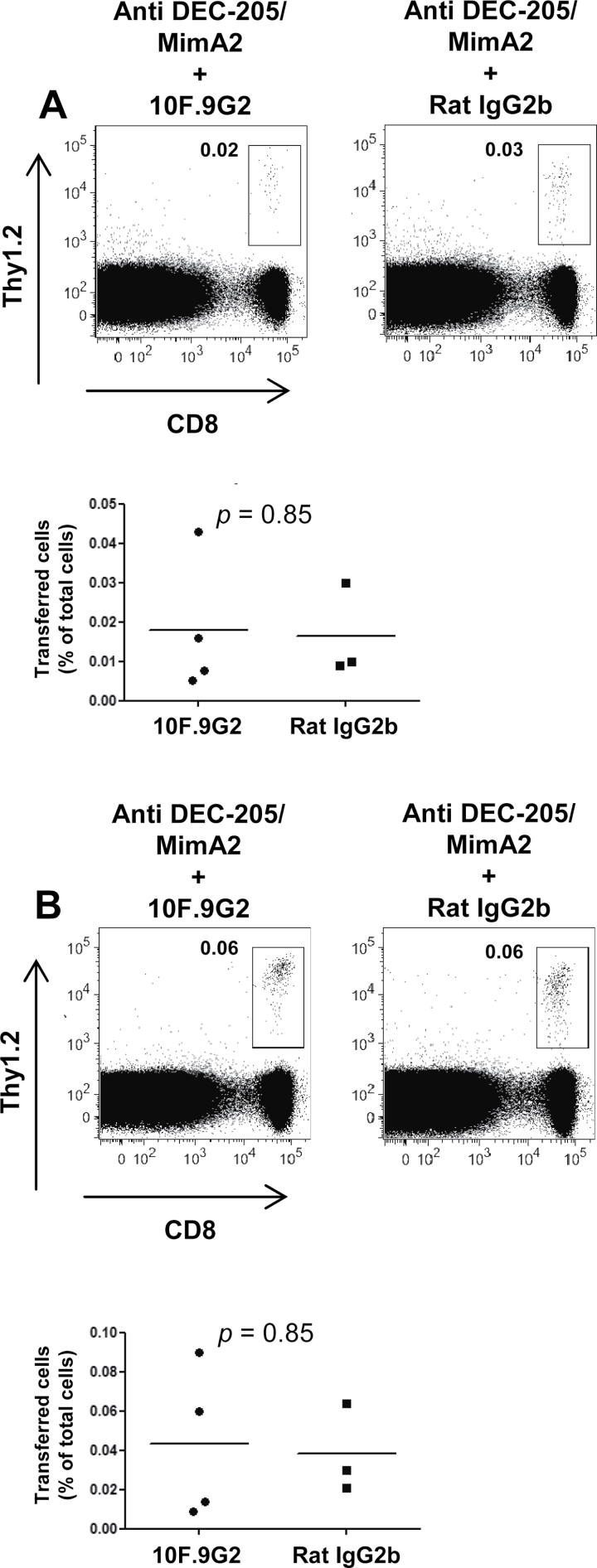
CD8⁺ T cells specific for islet-specific glucose-6-phosphatase catalytic subunit-related protein (IGRP) have been implicated in type 1 diabetes in both humans and non-obese diabetic (NOD) mice, in which T cells specific for IGRP₂₀₆₋₂₁₄ are highly prevalent. We sought to manipulate these pathogenic T cells by exploiting the ability of steady-state dendritic cells (DCs) to present antigens in a tolerogenic manner. The endocytic receptor DEC-205 was utilized to deliver an IGRP₂₀₆₋₂₁₄ mimotope to DCs in NOD mice, and the impact of this delivery on a polyclonal population of endogenous islet-reactive cognate T cells was determined. Assessment of islet-infiltrating CD8⁺ T cells showed a decrease in the percentage, and the absolute number, of endogenous IGRP₂₀₆₋₂₁₄-specific T cells when the mimotope was delivered to DCs, compared with delivery of a specificity control. Employing an adoptive transfer system, deletion of CD8⁺ T cells as a result of DEC-205-mediated antigen targeting was found to occur independently of programmed death-1 (PD-1) and its ligand (PD-L1), both often implicated in the regulation of peripheral T-cell tolerance. Given its promise for the manipulation of self-reactive polyclonal T cells demonstrated here, the distinctive characteristics of this antigen delivery system will be important to appreciate as its potential as an intervention for autoimmune diseases continues to be investigated.
Keywords: Autoimmunity; NOD mice; diabetes.
Figures
Similar articles
-
Selective delivery of beta cell antigen to dendritic cells in vivo leads to deletion and tolerance of autoreactive CD8+ T cells in NOD mice.Proc Natl Acad Sci U S A. 2008 Apr 29;105(17):6374-9. doi: 10.1073/pnas.0802644105. Epub 2008 Apr 22. Proc Natl Acad Sci U S A. 2008. PMID: 18430797 Free PMC article.
-
Efficient targeting of protein antigen to the dendritic cell receptor DEC-205 in the steady state leads to antigen presentation on major histocompatibility complex class I products and peripheral CD8+ T cell tolerance.J Exp Med. 2002 Dec 16;196(12):1627-38. doi: 10.1084/jem.20021598. J Exp Med. 2002. PMID: 12486105 Free PMC article.
-
Targeting DEC-205-DCIR2+ dendritic cells promotes immunological tolerance in proteolipid protein-induced experimental autoimmune encephalomyelitis.Mol Med. 2018 May 3;24(1):17. doi: 10.1186/s10020-018-0017-6. Mol Med. 2018. PMID: 30134798 Free PMC article.
-
Targeted antigen delivery to DEC-205⁺ dendritic cells for tolerogenic vaccination.Rev Diabet Stud. 2012 Winter;9(4):305-18. doi: 10.1900/RDS.2012.9.305. Epub 2012 Dec 28. Rev Diabet Stud. 2012. PMID: 23804268 Free PMC article. Review.
-
Applications of Antibody-Based Antigen Delivery Targeted to Dendritic Cells In Vivo.Antibodies (Basel). 2022 Jan 25;11(1):8. doi: 10.3390/antib11010008. Antibodies (Basel). 2022. PMID: 35225867 Free PMC article. Review.
Cited by
-
Dendritic Cells and Their Immunotherapeutic Potential for Treating Type 1 Diabetes.Int J Mol Sci. 2022 Apr 28;23(9):4885. doi: 10.3390/ijms23094885. Int J Mol Sci. 2022. PMID: 35563276 Free PMC article. Review.
-
Arrest in the Progression of Type 1 Diabetes at the Mid-Stage of Insulitic Autoimmunity Using an Autoantigen-Decorated All-trans Retinoic Acid and Transforming Growth Factor Beta-1 Single Microparticle Formulation.Front Immunol. 2021 Mar 8;12:586220. doi: 10.3389/fimmu.2021.586220. eCollection 2021. Front Immunol. 2021. PMID: 33763059 Free PMC article.
-
Targeting Dendritic Cells with Antigen-Delivering Antibodies for Amelioration of Autoimmunity in Animal Models of Multiple Sclerosis and Other Autoimmune Diseases.Antibodies (Basel). 2020 Jun 15;9(2):23. doi: 10.3390/antib9020023. Antibodies (Basel). 2020. PMID: 32549343 Free PMC article. Review.
-
Direct Delivery of Antigens to Dendritic Cells via Antibodies Specific for Endocytic Receptors as a Promising Strategy for Future Therapies.Vaccines (Basel). 2016 Mar 28;4(2):8. doi: 10.3390/vaccines4020008. Vaccines (Basel). 2016. PMID: 27043640 Free PMC article. Review.
-
Targeted immunomodulation using antigen-conjugated nanoparticles.Wiley Interdiscip Rev Nanomed Nanobiotechnol. 2014 May-Jun;6(3):298-315. doi: 10.1002/wnan.1263. Epub 2014 Mar 10. Wiley Interdiscip Rev Nanomed Nanobiotechnol. 2014. PMID: 24616452 Free PMC article. Review.
References
-
- Bonifaz L., Bonnyay D., Mahnke K., Rivera M., Nussenzweig M. C., Steinman R. M. 2002. Efficient targeting of protein antigen to the dendritic cell receptor DEC-205 in the steady state leads to antigen presentation on major histocompatibility complex class I products and peripheral CD8+ T cell tolerance. J. Exp. Med. 196:1627. - PMC - PubMed
-
- Hawiger D., Masilamani R. F., Bettelli E., Kuchroo V. K., Nussenzweig M. C. 2004. Immunological unresponsiveness characterized by increased expression of CD5 on peripheral T cells induced by dendritic cells in vivo . Immunity 20:695. - PubMed
Publication types
MeSH terms
Substances
Grants and funding
LinkOut - more resources
Full Text Sources
Other Literature Sources
Molecular Biology Databases
Research Materials
