

Ammonia binding to the oxygen-evolving complex of photosystem II identifies the solvent-exchangeable oxygen bridge (μ-oxo) of the manganese tetramer
- PMID: 24023065
- PMCID: PMC3785721
- DOI: 10.1073/pnas.1304334110
Ammonia binding to the oxygen-evolving complex of photosystem II identifies the solvent-exchangeable oxygen bridge (μ-oxo) of the manganese tetramer
Abstract
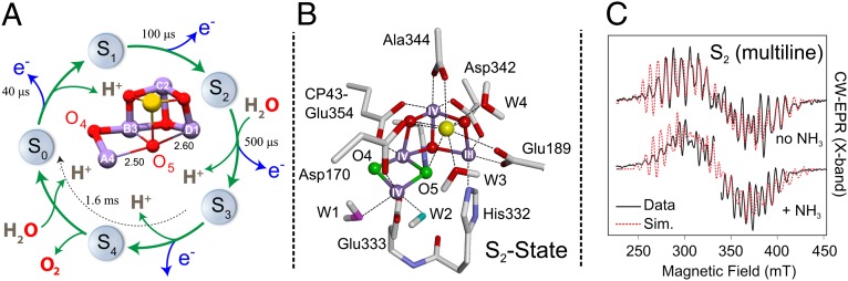
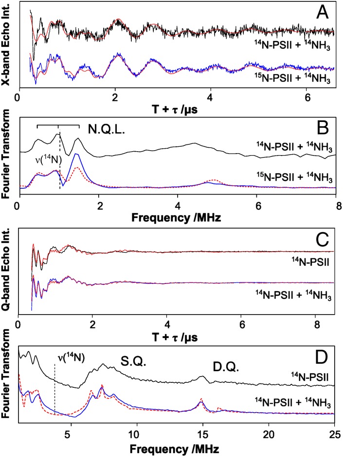
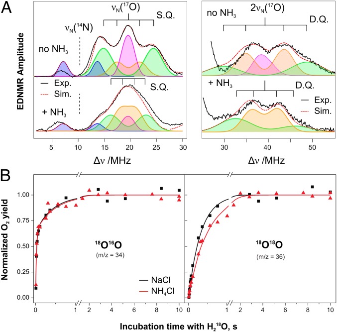
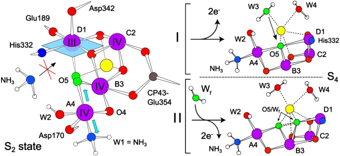
The assignment of the two substrate water sites of the tetra-manganese penta-oxygen calcium (Mn4O5Ca) cluster of photosystem II is essential for the elucidation of the mechanism of biological O-O bond formation and the subsequent design of bio-inspired water-splitting catalysts. We recently demonstrated using pulsed EPR spectroscopy that one of the five oxygen bridges (μ-oxo) exchanges unusually rapidly with bulk water and is thus a likely candidate for one of the substrates. Ammonia, a water analog, was previously shown to bind to the Mn4O5Ca cluster, potentially displacing a water/substrate ligand [Britt RD, et al. (1989) J Am Chem Soc 111(10):3522-3532]. Here we show by a combination of EPR and time-resolved membrane inlet mass spectrometry that the binding of ammonia perturbs the exchangeable μ-oxo bridge without drastically altering the binding/exchange kinetics of the two substrates. In combination with broken-symmetry density functional theory, our results show that (i) the exchangable μ-oxo bridge is O5 {using the labeling of the current crystal structure [Umena Y, et al. (2011) Nature 473(7345):55-60]}; (ii) ammonia displaces a water ligand to the outer manganese (MnA4-W1); and (iii) as W1 is trans to O5, ammonia binding elongates the MnA4-O5 bond, leading to the perturbation of the μ-oxo bridge resonance and to a small change in the water exchange rates. These experimental results support O-O bond formation between O5 and possibly an oxyl radical as proposed by Siegbahn and exclude W1 as the second substrate water.
Keywords: Mn cluster; OEC; PSII; water oxidizing complex; water-oxidation.
Conflict of interest statement
The authors declare no conflict of interest.
Figures
Similar articles
-
Detection of the water-binding sites of the oxygen-evolving complex of Photosystem II using W-band 17O electron-electron double resonance-detected NMR spectroscopy.J Am Chem Soc. 2012 Oct 10;134(40):16619-34. doi: 10.1021/ja3053267. Epub 2012 Sep 27. J Am Chem Soc. 2012. PMID: 22937979
-
Mono-manganese mechanism of the photosystem II water splitting reaction by a unique Mn4Ca cluster.Biochim Biophys Acta. 2007 Jun;1767(6):484-92. doi: 10.1016/j.bbabio.2007.03.012. Epub 2007 Apr 4. Biochim Biophys Acta. 2007. PMID: 17490604 Review.
-
The O2-Evolving Complex of Photosystem II: Recent Insights from Quantum Mechanics/Molecular Mechanics (QM/MM), Extended X-ray Absorption Fine Structure (EXAFS), and Femtosecond X-ray Crystallography Data.Acc Chem Res. 2017 Jan 17;50(1):41-48. doi: 10.1021/acs.accounts.6b00405. Epub 2016 Dec 21. Acc Chem Res. 2017. PMID: 28001034 Review.
-
EPR-ENDOR characterization of (17O, 1H, 2H) water in manganese catalase and its relevance to the oxygen-evolving complex of photosystem II.J Am Chem Soc. 2012 Jan 25;134(3):1504-12. doi: 10.1021/ja203465y. Epub 2012 Jan 9. J Am Chem Soc. 2012. PMID: 22142421 Free PMC article.
-
Structure, ligands and substrate coordination of the oxygen-evolving complex of photosystem II in the S2 state: a combined EPR and DFT study.Phys Chem Chem Phys. 2014 Jun 28;16(24):11877-92. doi: 10.1039/c3cp55017f. Phys Chem Chem Phys. 2014. PMID: 24525937
Cited by
-
CaMn3IV O4 Cubane Models of the Oxygen-Evolving Complex: Spin Ground States S<9/2 and the Effect of Oxo Protonation.Angew Chem Int Ed Engl. 2021 Aug 2;60(32):17671-17679. doi: 10.1002/anie.202105303. Epub 2021 Jul 1. Angew Chem Int Ed Engl. 2021. PMID: 34042234 Free PMC article.
-
Removal of Ca(2+) from the Oxygen-Evolving Complex in Photosystem II Has Minimal Effect on the Mn4O5 Core Structure: A Polarized Mn X-ray Absorption Spectroscopy Study.J Phys Chem B. 2015 Oct 29;119(43):13742-54. doi: 10.1021/acs.jpcb.5b03559. Epub 2015 Jun 3. J Phys Chem B. 2015. PMID: 25989608 Free PMC article.
-
Probing substrate water access through the O1 channel of Photosystem II by single site mutations and membrane inlet mass spectrometry.Photosynth Res. 2025 Apr 22;163(3):28. doi: 10.1007/s11120-025-01147-4. Photosynth Res. 2025. PMID: 40263146 Free PMC article.
-
Functional Water Networks in Fully Hydrated Photosystem II.J Am Chem Soc. 2022 Dec 7;144(48):22035-22050. doi: 10.1021/jacs.2c09121. Epub 2022 Nov 22. J Am Chem Soc. 2022. PMID: 36413491 Free PMC article.
-
Plasma Membrane-Type Aquaporins from Marine Diatoms Function as CO2/NH3 Channels and Provide Photoprotection.Plant Physiol. 2018 Sep;178(1):345-357. doi: 10.1104/pp.18.00453. Epub 2018 Aug 3. Plant Physiol. 2018. PMID: 30076224 Free PMC article.
References
-
- Umena Y, Kawakami K, Shen J-R, Kamiya N. Crystal structure of oxygen-evolving photosystem II at a resolution of 1.9 Å. Nature. 2011;473(7345):55–60. - PubMed
-
- Ferreira KN, Iverson TM, Maghlaoui K, Barber J, Iwata S. Architecture of the photosynthetic oxygen-evolving center. Science. 2004;303(5665):1831–1838. - PubMed
-
- Loll B, Kern J, Saenger W, Zouni A, Biesiadka J. Towards complete cofactor arrangement in the 3.0 Å resolution structure of photosystem II. Nature. 2005;438(7070):1040–1044. - PubMed
-
- Kok B, Forbush B, McGloin M. Cooperation of charges in photosynthetic O2 evolution-I. A linear four step mechanism. Photochem Photobiol. 1970;11(6):457–475. - PubMed
Publication types
MeSH terms
Substances
LinkOut - more resources
Full Text Sources
Other Literature Sources
Research Materials
Miscellaneous
