. 2013 Oct 8;110(41):16568-73.
doi: 10.1073/pnas.1308755110. Epub 2013 Sep 10.

HIF1α is required for osteoclast activation by estrogen deficiency in postmenopausal osteoporosis

Affiliations

HIF1α is required for osteoclast activation by estrogen deficiency in postmenopausal osteoporosis

Yoshiteru Miyauchi et al. Proc Natl Acad Sci U S A. .

Abstract

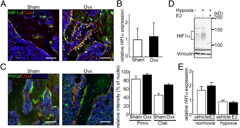
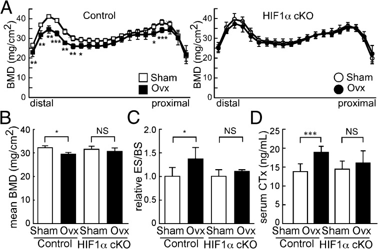
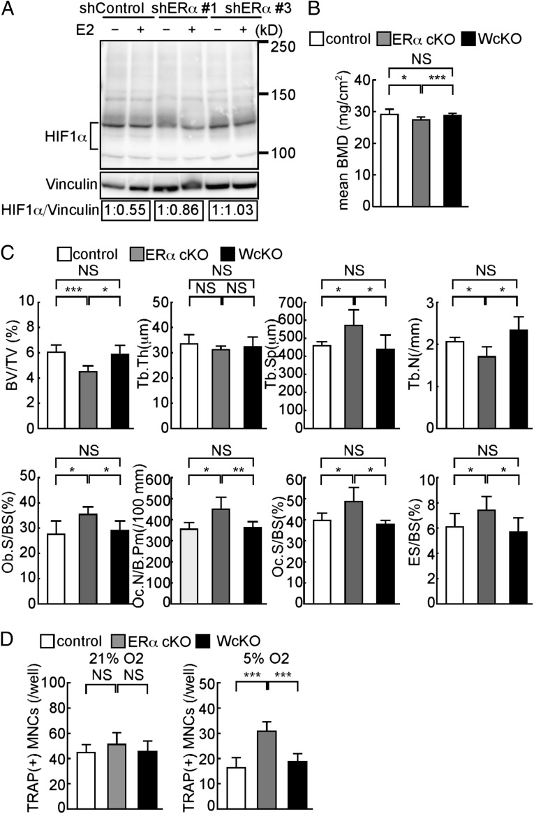
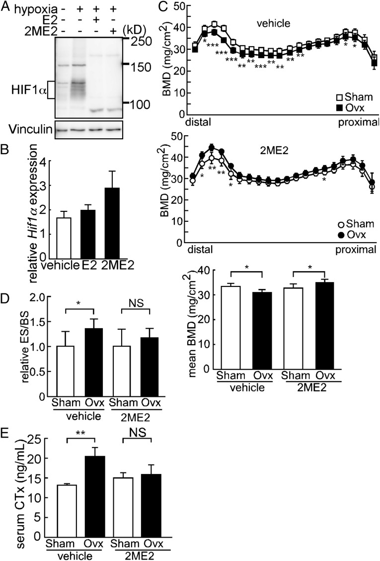
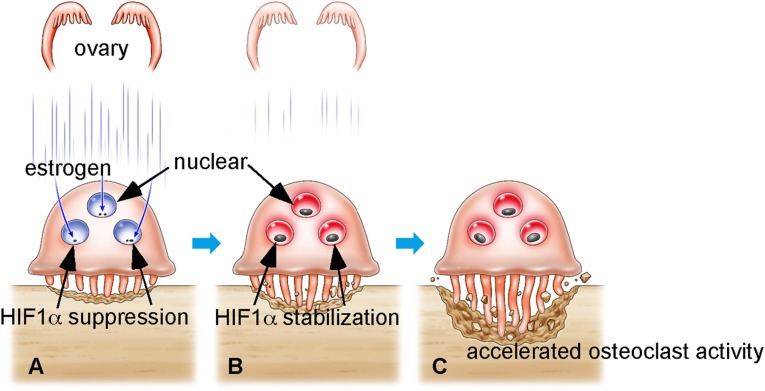
In women, estrogen deficiency after menopause frequently accelerates osteoclastic bone resorption, leading to osteoporosis, the most common skeletal disorder. However, mechanisms underlying osteoporosis resulting from estrogen deficiency remain largely unknown. Here we show that in bone-resorbing osteoclasts, estrogen-dependent destabilization of hypoxia-inducible factor 1 alpha (HIF1α), which is unstable in the presence of oxygen, plays a pivotal role in promoting bone loss in estrogen-deficient conditions. In vitro, HIF1α was destabilized by estrogen treatment even in hypoxic conditions, and estrogen loss in ovariectomized (Ovx) mice stabilized HIF1α in osteoclasts and promoted their activation and subsequent bone loss in vivo. Osteoclast-specific HIF1α inactivation antagonized bone loss in Ovx mice and osteoclast-specific estrogen receptor alpha deficient mice, both models of estrogen-deficient osteoporosis. Oral administration of a HIF1α inhibitor protected Ovx mice from osteoclast activation and bone loss. Thus, HIF1α represents a promising therapeutic target in osteoporosis.


Conflict of interest statement

The authors declare no conflict of interest.

Figures

Fig. 1.
Fig. 1.
Estrogen destabilizes HIF1α in vivo and in vitro. (A) Immunofluorescence for HIF1α (green) and Ctsk (red) in bone sections from Sham or Ovx mice. Nuclei are stained with TOTO-3 (blue). *, HIF1α/Ctsk double-positive cells. (B) Hif1α mRNA levels in c-Fms+Mac1high osteoclasts sorted from Sham or Ovx mouse bone marrow as determined by quantitative PCR. Cells were prepared 5 wk after surgery. (C, Left) Immunofluorescent detection of hypoxic areas (Pimo, green) and Ctsk (red) on bone sections from Sham or Ovx mice. Nuclei are stained with TOTO-3 (blue). (Scale bar, 100 μm.) (C, Right) Signal intensities of Pimo and Ctsk shown as mean values relative to TOTO-3 ± SD (n = 3). (D) Western blot analysis of HIF1α in Raw264.7 cells cultured with or without E2. (E) Hif1α mRNA levels in Raw264.7 cells as determined by quantitative PCR in the presence or absence of E2 under normoxic or hypoxic conditions. Data in B and E represent mean Hif1α expression relative to Actb ± SD (n = 3).
Fig. 2.
Fig. 2.
HIF1α cKO mice are resistant to ovariectomy-induced bone loss and osteoclast activation. (A and B) BMD of femurs divided equally longitudinally from Sham or Ovx control (Hif1αf/f) and HIF1α cKO (CtskCre/+; Hif1αf/f) mice. (C) ES/BS of femurs from sham-operated or ovariectomized control and HIF1α cKO mice. (D) Levels of serum CTx, a marker of bone resorption, in control and HIF1α cKO mice. Analysis was undertaken 5 wk after Ovx or Sham surgery. All data are means ± SD (n = 5). *P < 0.05; **P < 0.01; ***P < 0.005; NS, not significant.
Fig. 3.
Fig. 3.
HIF1α deletion rescues bone phenotypes seen in ERα cKO mice in vivo. (A) Raw264.7 cells transfected with shRNA targeting ERα (shERα) or nontarget control shRNA (shControl) and treated with (+) or without (−) E2. HIF1α protein levels determined by immunoblot were quantified by densitometry and are shown as values relative to E2(−). (B) BMD of femurs divided equally longitudinally from control (CtskCre/+), ERα cKO (CtskCre/+; ERαf/f), and WcKO (CtskCre/+; Hif1αf/f; ERαf/f) mice. (C) Bone histomorphometrical analysis of femurs from control, ERα cKO, and WcKO female mice. (D) Osteoclast formation, as evidenced by TRAP positivity, of osteoclast progenitor cells from control, ERα cKO, and WcKO mouse bone marrow under normoxic (21%) or hypoxic (5%) conditions during the course of RANKL-stimulated differentiation. Data (BD) represent the mean value of the indicated parameter ± SD (*P < 0.05; ***P < 0.005; NS, not significant; n = 5). White, gray, and black bars represent control, ERα cKO, and WcKO mice, respectively.
Fig. 4.
Fig. 4.
Pharmacological HIF1α ablation antagonizes Ovx-induced bone loss and osteoclast activation. (A) Suppression of HIF1α protein expression in Raw264.7 cells by E2 or 2ME2 in hypoxic conditions. (B) Effects of E2 or 2ME2 on Hif1α mRNA levels in Raw264.7 cells. Data represent mean Hif1α expression relative to that of Actb ± SD (n = 5). (C, Top, Middle, and Bottom) BMD of femurs from Sham or Ovx treated with vehicle or 2ME2. (D) ES/BS of femurs from Sham or Ovx treated with vehicle or 2ME2. (E) Serum CTx of Sham or Ovx mice treated with vehicle or 2ME2. Error bars indicate means ± SD (n = 5). *P < 0.05; **P < 0.01; ***P < 0.005; NS, not significant.
Fig. 5.
Fig. 5.
Schematic model of postmenopausal osteoporosis. (A) Osteoclasts are localized in the endosteal zone, where oxygen levels are low. Even in hypoxic conditions, HIFα protein is continuously destabilized by estrogen in osteoclasts. (B) After menopause or Ovx, HIFα is stabilized in osteoclasts due to estrogen depletion. (C) HIFα stabilization in osteoclasts promotes osteoclast activation, which promotes reduced bone mass and osteoporosis.

References

    1. Delmas PD. Treatment of postmenopausal osteoporosis. Lancet. 2002;359(9322):2018–2026. - PubMed
    1. Rodan GA, Martin TJ. Therapeutic approaches to bone diseases. Science. 2000;289(5484):1508–1514. - PubMed
    1. Nelson ER, Wardell SE, McDonnell DP. The molecular mechanisms underlying the pharmacological actions of estrogens, SERMs and oxysterols: Implications for the treatment and prevention of osteoporosis. Bone. 2013;53(1):42–50. - PMC - PubMed
    1. Riggs BL, Hartmann LC. Selective estrogen-receptor modulators—Mechanisms of action and application to clinical practice. N Engl J Med. 2003;348(7):618–629. - PubMed
    1. Couse JF, Korach KS. Estrogen receptor null mice: What have we learned and where will they lead us? Endocr Rev. 1999;20(3):358–417. - PubMed

Publication types

MeSH terms