. 2013 Sep 10;4(5):e00650-13.
doi: 10.1128/mBio.00650-13.

Engineering a replication-competent, propagation-defective Middle East respiratory syndrome coronavirus as a vaccine candidate

Affiliations

Engineering a replication-competent, propagation-defective Middle East respiratory syndrome coronavirus as a vaccine candidate

Fernando Almazán et al. mBio. .

Abstract

Middle East respiratory syndrome coronavirus (MERS-CoV) is an emerging coronavirus infecting humans that is associated with acute pneumonia, occasional renal failure, and a high mortality rate and is considered a threat to public health. The construction of a full-length infectious cDNA clone of the MERS-CoV genome in a bacterial artificial chromosome is reported here, providing a reverse genetics system to study the molecular biology of the virus and to develop attenuated viruses as vaccine candidates. Following transfection with the cDNA clone, infectious virus was rescued in both Vero A66 and Huh-7 cells. Recombinant MERS-CoVs (rMERS-CoVs) lacking the accessory genes 3, 4a, 4b, and 5 were successfully rescued from cDNA clones with these genes deleted. The mutant viruses presented growth kinetics similar to those of the wild-type virus, indicating that accessory genes were not essential for MERS-CoV replication in cell cultures. In contrast, an engineered mutant virus lacking the structural E protein (rMERS-CoV-ΔE) was not successfully rescued, since viral infectivity was lost at early passages. Interestingly, the rMERS-CoV-ΔE genome replicated after cDNA clone was transfected into cells. The infectious virus was rescued and propagated in cells expressing the E protein in trans, indicating that this virus was replication competent and propagation defective. Therefore, the rMERS-CoV-ΔE mutant virus is potentially a safe and promising vaccine candidate to prevent MERS-CoV infection.

Importance: Since the emergence of MERS-CoV in the Arabian Peninsula during the summer of 2012, it has already spread to 10 different countries, infecting around 94 persons and showing a mortality rate higher than 50%. This article describes the development of the first reverse genetics system for MERS-CoV, based on the construction of an infectious cDNA clone inserted into a bacterial artificial chromosome. Using this system, a collection of rMERS-CoV deletion mutants has been generated. Interestingly, one of the mutants with the E gene deleted was a replication-competent, propagation-defective virus that could only be grown in the laboratory by providing E protein in trans, whereas it would only survive a single virus infection cycle in vivo. This virus constitutes a vaccine candidate that may represent a balance between safety and efficacy for the induction of mucosal immunity, which is needed to prevent MERS-CoV infection.


Figures

FIG 1
FIG 1
Assembly of a MERS-CoV full-length cDNA clone as a BAC. (A) Genome organization of the MERS-CoV-EMC12 strain. Viral genes (ORF 1a, ORF 1b, S, 3, 4a, 4b, 5, E, M, and N) are illustrated by boxes in this genome scheme. The relevant restriction sites used for the assembly of the infectious cDNA clone and their genomic positions (first nucleotide of the recognition sequence) are indicated. L, leader sequence; UTR, untranslated region; An, poly(A) tail. (B) Schematic representation of pBAC-MERS-5′3′. Relevant restriction sites, the CMV transcription start, the HDV ribozyme (Rz), and the BGH termination and polyadenylation sequences (BGH) are shown. (C) Strategy to assemble the MERS-CoV infectious cDNA clone. Four overlapping DNA fragments (MERS-1 to MERS-4), generated by chemical synthesis, were sequentially cloned into the plasmid pBAC-MERS-5′3′ to generate the MERS-CoV infectious cDNA clone (pBAC-MERSFL). Relevant restriction sites and the genetic marker (T to C) introduced at position 20,761 to abrogate the SwaI restriction site at position 20,760 are indicated. Acronyms for viral genes and regulatory elements are as described for panels A and B.
FIG 2
FIG 2
Identification of the virus recovered from the cDNA clone. Vero A66 and Huh-7 cells were mock infected or infected with the rMERS-CoV rescued in these cell lines at an MOI of 0.001 PFU/cell. The induction of syncytium formation (CPE) and N protein expression were analyzed 48 h.p.i. by light microscopy and indirect immunofluorescence assay (IFA), respectively. Pictures were taken with a 40× objective.
FIG 3
FIG 3
Rescue and growth kinetics of rMERS-CoV deletion mutants. (A) Genetic structure of rMERS-CoV-∆3, rMERS-CoV-∆4ab, and rMERS-CoV-∆5 deletion mutants. TRSs and viral genes are depicted as boxes. The genomic positions of the deletions introduced (gray boxes) are indicated; the numbers correspond to the last and first nondeleted nucleotide in each case. Acronyms for viral genes are as defined in the legend to Fig. 1. (B) Growth kinetics of the deletion mutants. Huh-7 cells were infected at an MOI of 0.001 PFU/cell with rMERS-CoV-∆3, rMERS-CoV-∆4ab, rMERS-CoV-∆5, or the wild-type virus (rMERS-CoV), and at the indicated times postinfection, virus titers were determined by plaque assay on Huh-7 cells. Error bars represent standard deviations of the mean from three experiments.
FIG 4
FIG 4
Rescue of rMERS-CoV-ΔE. (A) Genetic structure of rMERS-CoV-ΔE. TRSs and viral genes are illustrated as boxes. The genomic position of the introduced deletion (gray box) is indicated; the numbers correspond to the last and first nondeleted nucleotides. Acronyms for viral genes are as defined in the legend to Fig. 1. (B) Identification of the recovered viruses by immunofluorescence microscopy. Huh-7 cells were mock infected (MOCK) or infected with rMERS-CoV-ΔE and rMERS-CoV viruses from passage 0, and the expression of viral proteins E and N was analyzed 48 h.p.i. by immunofluorescence microscopy using specific antibodies.
FIG 5
FIG 5
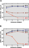
Rescue of rMERS-CoV-ΔE in cells expressing E protein in trans. (A) Virus rescue. After Huh-7 cells expressing (E+) or not expressing (E) the E protein in trans were transfected with plasmids pBAC-MERS-ΔE and pBAC-MERSFL, cell culture supernatants were serially passaged 3 times on fresh E+ and E cells every 72 h.p.i., and the virus titers of the rescued rMERS-CoV (WT) and rMERS-CoV-ΔE (ΔE) were determined by limiting dilution. The black dashed line represents the detection threshold of the virus titration assay (50 TCID50/ml). Error bars represent standard deviations of the means from three experiments. (B) Viral gRNA analysis. The levels of viral gRNA in E+ and E cells infected with either rMERS-CoV (WT) or rMERS-CoV-ΔE (ΔE) were analyzed at each passage. Total RNA was extracted and analyzed by RT-qPCR. gRNA levels were normalized by 18S rRNA levels. Error bars represent standard deviations of the means from three experiments.
FIG 6
FIG 6
Analysis of replication and transcription levels in rMERS-CoV-ΔE-infected cells. Huh-7 cells were infected with rMERS-CoV-ΔE and rMERS-CoV at an MOI of 0.001 TCDI50/ml, and at 5 h.p.i., the levels of gRNA and sgmRNA N were evaluated by RT-qPCR. Both gRNA and sgmRNA N levels were normalized by 18S rRNA levels. In addition, sgmRNA N levels were made relative to gRNA levels. Error bars represent standard deviations of the means from three experiments.

Similar articles

Cited by

References

    1. Danielsson N, Catchpole M, ECDC Internal Response Team 2012. Novel coronavirus associated with severe respiratory disease: case definition and public health measures. Euro Surveill. 17:20282. - PubMed
    1. Zaki AM, van Boheemen S, Bestebroer TM, Osterhaus AD, Fouchier RA. 2012. Isolation of a novel coronavirus from a man with pneumonia in Saudi Arabia. N. Engl. J. Med. 367:1814–1820 - PubMed
    1. MMWR 2013. Update: severe respiratory illness associated with Middle East respiratory syndrome coronavirus (MERS-CoV)—worldwide, 2012–2013. Morb. Mortal. Wkly. Rep. 62:480–483 - PMC - PubMed
    1. de Groot RJ, Baker SC, Baric RS, Brown CS, Drosten C, Enjuanes L, Fouchier RA, Galiano M, Gorbalenya AE, Memish ZA, Perlman S, Poon LL, Snijder EJ, Stephens GM, Woo PC, Zaki AM, Zambon M, Ziebuhr J. 2013. Middle East respiratory syndrome coronavirus (MERS-CoV): announcement of the Coronavirus Study Group. J. Virol. 87:7790–7792 - PMC - PubMed
    1. Annan A, Baldwin HJ, Corman VM, Klose SM, Owusu M, Nkrumah EE, Badu EK, Anti P, Agbenyega O, Meyer B, Oppong S, Sarkodie YA, Kalko EK, Lina PH, Godlevska EV, Reusken C, Seebens A, Gloza-Rausch F, Vallo P, Tschapka M, Drosten C, Drexler JF. 2013. Human betacoronavirus 2c EMC/2012-related viruses in bats, Ghana and Europe. Emerg. Infect. Dis. 19:456–459 - PMC - PubMed

Publication types