. 2013 Sep 2;8(9):e72011.
doi: 10.1371/journal.pone.0072011. eCollection 2013.

M1 muscarinic receptor activation mediates cell death in M1-HEK293 cells

Affiliations

M1 muscarinic receptor activation mediates cell death in M1-HEK293 cells

E Scott Graham et al. PLoS One. .

Abstract

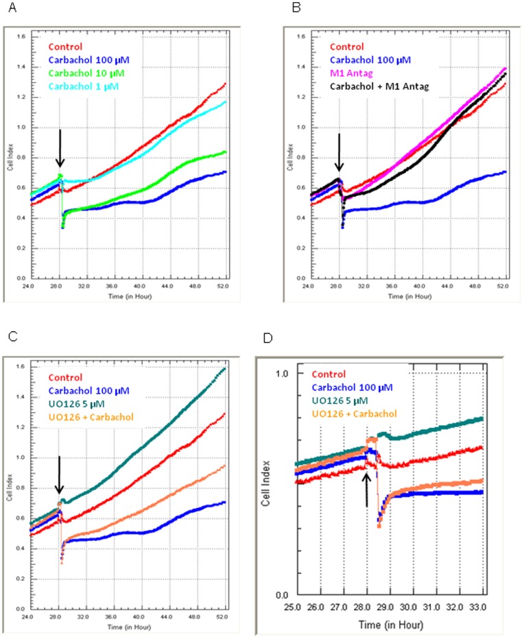
HEK293 cells have been used extensively to generate stable cell lines to study G protein-coupled receptors, such as muscarinic acetylcholine receptors (mAChRs). The activation of M1 mAChRs in various cell types in vitro has been shown to be protective. To further investigate M1 mAChR-mediated cell survival, we generated stable HEK293 cell-lines expressing the human M1 mAChR. M1 mAChRs were efficiently expressed at the cell surface and efficiently internalised within 1 h by carbachol. Carbachol also induced early signalling cascades similar to previous reports. Thus, ectopically expressed M1 receptors behaved in a similar fashion to the native receptor over short time periods of analysis. However, substantial cell death was observed in HEK293-M1 cells within 24 h after carbachol application. Death was only observed in HEK cells expressing M1 receptors and fully blocked by M1 antagonists. M1 mAChR-stimulation mediated prolonged activation of the MEK-ERK pathway and resulted in prolonged induction of the transcription factor EGR-1 (>24 h). Blockade of ERK signalling with U0126 did not reduce M1 mAChR-mediated cell-death significantly but inhibited the acute induction of EGR-1. We investigated the time-course of cell death using time-lapse microscopy and xCELLigence technology. Both revealed the M1 mAChR cytotoxicity occurs within several hours of M1 activation. The xCELLigence assay also confirmed that the ERK pathway was not involved in cell-death. Interestingly, the MEK blocker did reduce carbachol-mediated cleaved caspase 3 expression in HEK293-M1 cells. The HEK293 cell line is a widely used pharmacological tool for studying G-protein coupled receptors, including mAChRs. Our results highlight the importance of investigating the longer term fate of these cells in short term signalling studies. Identifying how and why activation of the M1 mAChR signals apoptosis in these cells may lead to a better understanding of how mAChRs regulate cell-fate decisions.


Conflict of interest statement

Competing Interests: The authors have declared that no competing interests exist.

Figures

Figure 1
Figure 1. M1 mAChR expression and localisation in transfected HEK293 cells.
(A) PCR was conducted to assess M1 expression by vector-transfected (HEK293-Vec) and M1 transfected HEK293 cells (HEK293-M1) using primers for M1 mAChR. The integrity of cDNA samples was confirmed using GAPDH. Samples are as follows; HEK293-Vec cDNA in lane 1, HEK293-Vec -RT/RNA in lane 2, HEK293-M1 cDNA in lane 3, and HEK293-M1–RT/RNA in lane 4. Samples are representative of those used in subsequent functional studies. (B) Immunocytochemical localisation of M1 receptors in HEK293-M1 cells using anti-HA.11 antibody (middle panel) and M1 antibody (right-hand panel). Left panel shows no primary control Image. (C) Analysis of M1 receptor cell-surface expression and internalization by carbachol. Cell surface receptors were live-labeled (see methods) using the HA.11antibody prior to stimulation with water-control or carbachol treatment. The M1 mAChRs were typically localised at the plasma membrane after water treatment, but after 1 h carbachol treatment for the M1 mAChRs were internalised, as shown by the increased punctate cytoplasmic staining (arrows) and reduced staining intensity on the cell surfaces. Scale bar: 50 µm. Data are representative of at least three independent experiments. (D) shows the time-course of M1 receptor internalization after carbachol addition using the granularity assay in Metamorph to measure internalized receptors (as intracellular granules). The graph shows that 5–60 minutes after carbachol addition there is internalization of M1 receptors.
Figure 2
Figure 2. pERK1/2 and EGR-1 induction in transfected HEK293-M1 cells by carbachol.
Expression of pERK1/2 and EGR-1 after short term carbachol treatment in HEK293-Vec cells and HEK293-M1 cells. Cells were stimulated for 10 minutes and 1 h for pERK and 1 h for EGR-1 expression with 100 µM carbachol or vehicle. Data are representative of at least three independent experiments. Scale bar: 50 µm.
Figure 3
Figure 3. Acute activation of EGR-1 by M1 receptors is ERK dependent in HEK293-M1 cells.
HEK293-M1 cells were pretreated with 5 µM UO126 (or vehicle) for 30 mins prior to the addition of 100 µM carbachol for the activation of pERK (10 mins) and EGR-1 (1 h). Data are representative of at least three independent experiments. Scale bar: 50 µM. Graphs show quantification of the induction and reversal of pERK and EGR-1 with U0126.
Figure 4
Figure 4. Chronic M1 mAChRs activation induces HEK293-M1 cell death.
(A) Hoechst staining of HEK293-M1 and HEK293-Vec treated with water control (vehicle) or 100 µM carbachol for 24 h. The arrows point to condensed (possibly apoptotic) nuclei that were present in carbachol-treated HEK293-M1 cells. Note the substantial cell loss in the M1 expressing HEK cells, but not in the vector transfected HEK cells. Scale bar: 25 µm. (B) The extent of cell loss was investigated further using the MEK-inhibitor UO126. Hoechst stained cells were quantified using Discovery-1 and Metamorph analysis. Statistical significance: *** denotes p<0.001 compared with all other conditions. Note that UO126 did not significantly affect the carbachol response (ns) (C) HEK293-M1 cell counts across a range of 24 h carbachol treatments also showed that cell death occurred in a concentration-dependent manner. Data are representative of at least three independent experiments.
Figure 5
Figure 5. Analysis of carbachol mediated cell death using xCELLigence technology.
HEK293-M1 cells were stimulated with various concentrations of carbachol (A), in the presence (or absence) of a M1 selective antagonist (B), and with 5 µM UO126 (C and D). Panel D shows the period immediately following drug addition to highlight the immediacy of the carbachol response. In each graph the black arrow shows the time point the respective drugs were added. Cellular measurements are made continuously in real time, where the level of total cell adhesion is represented as Cell Index. Higher Cell Index levels represent more adhesion, whereas a reduction in Cell Index represents a loss of adhesion. In all four graphs the red and blue Cell Index curves are the same and represent vehicle-treated and 100 µM carbachol-treated HEK293-M1 cells. The reduction in adhesion mediated by carbachol is immediate, concentration-dependent, blocked by M1 antagonist, but not blocked by UO126 (MEK blocker). xCELLigence data show representative Cell Index curves of at least six independent experiments.
Figure 6
Figure 6. M1 mAChR activation induces cleaved-caspase 3 but does not induce changes in proliferation.
HEK293-M1 and HEK293-Vec cells were stimulated across a 48 hour time course (2, 4, 8, 24, and 48 hours) with 100 µM carbachol (A). Induction of cleaved caspase-3 was evident following carbachol but only in the HEK293-M1 (M1) expressing cells. The induction was statistically significant at 24 hours and by 48 hours almost 50% of the cells expressed cleaved caspase 3. In contrast, there was no expression of cleaved caspase 3 in HEK-Vector (Vec) expressing cells. (B. To determine whether this expression was M1-mediated and MEK-mediated HEK293-M1 cells were incubated with the M1 antagonist MT7 or the MEK blocker U0126 and then challenged with carbachol. Induction of cleaved caspase 3 by carbachol was strongly inhibited by both drugs indicating that it was M1- and MEK-mediated. C. Photomicrographs of cleaved caspase 3 in HEK293-M1 cells 48 hours after vehicle (left image) or carbachol (right image) addition showing strong induction of cleaved caspase 3 in carbachol treated cells. (D) HEK293-Vec cells and HEK293-M1 cells were stimulated with 100 µM carbachol or vehicle for 24 hours, where BrdU was included during the final hour of treatment. BrdU incorporation revealed that the carbachol mediated reduction in cell numbers was not as a consequence of influencing proliferation. BrdU positive cells are as a percentage of the total cells counted to represent percentage proliferating cells. Data are representative of at least three independent experiments.
Figure 7
Figure 7. Activation of pERK and EGR-1 is still evident 24 hours following initial carbachol treatment.
HEK293-M1 cells were stimulated for 24 h with vehicle or 100 µM carbachol as indicated. Then the cells were fixed and stained for pERK and EGR1. Note the high uniform activation of pERK and EGR-1. The lower panels show the effect of pretreatment (30 minutes) with 5 µM UO126, which shows that the aberrant signalling present at 24 h is independent of the MEK signalling pathway. Graphs show quantification of the induction and reversal of pERK and EGR-1 with U0126.
Figure 8
Figure 8. Western blots of pERK (A) and EGR-1 (B) at 10 min, 1 hr and 24 hours after Carbachol +/− U0126.
A, pERK Western showing ERK phosphorylation at 10-min and 24 hours after addition of carbachol to HEK293-M1 cells. The MEK blocker U0126 completely inhibited this ERK phosphorylation. B, EGR-1 induction at 1 hour and 24 hours after addition of carbachol to HEK293-M1 cells. The MEK blocker U0126 completely inhibited this EGR-1 induction.
Figure 9
Figure 9. Schematic diagram showing that M1-induced cell death involved two components.
The major component of death was ERK-independent and occurred within hours of carbachol activation of the cells. This pathway was not blocked by the MEK inhibitor UO126 and occurred prior to activation of cleaved caspase-3. In contrast the minor component, which was slower and more progressive, was blocked by UO126 and associated with cleaved caspase-3 expression. Both pathways were fully blocked by the M1 antagonist and only occurred in HEK-M1 cells stably expressing M1 receptors.

Similar articles

Cited by

References

    1. Wess J (2004) Muscarinic acetylcholine receptor knockout mice: novel phenotypes and clinical implications. Annu Rev Pharmacol Toxicol 44: 423–450. - PubMed
    1. Dragunow M (2008) M3 muscarinic receptors as targets for drug development in neurodegenerative disorders. Nat Rev Drug Discov 7: 1 p following p185. - PubMed
    1. Yan GM, Lin SZ, Irwin RP, Paul SM (1995) Activation of muscarinic cholinergic receptors blocks apoptosis of cultured cerebellar granule neurons. Mol Pharmacol 47: 248–257. - PubMed
    1. Giordano G, Li L, White CC, Farin FM, Wilkerson HW, et al. (2009) Muscarinic receptors prevent oxidative stress-mediated apoptosis induced by domoic acid in mouse cerebellar granule cells. J Neurochem 109: 525–538. - PMC - PubMed
    1. De Sarno P, Shestopal SA, King TD, Zmijewska A, Song L, et al. (2003) Muscarinic receptor activation protects cells from apoptotic effects of DNA damage, oxidative stress, and mitochondrial inhibition. J Biol Chem 278: 11086–11093. - PMC - PubMed

Publication types