. 2013 Sep 2;8(9):e72120.
doi: 10.1371/journal.pone.0072120. eCollection 2013.

Metformin inhibits angiotensin II-induced differentiation of cardiac fibroblasts into myofibroblasts

Affiliations

Metformin inhibits angiotensin II-induced differentiation of cardiac fibroblasts into myofibroblasts

Jian Bai et al. PLoS One. .

Abstract

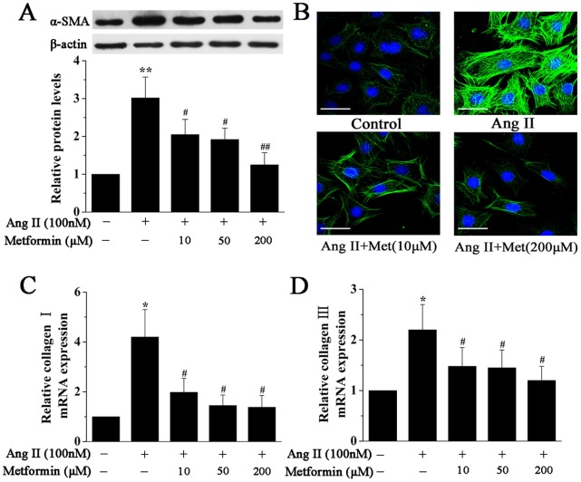
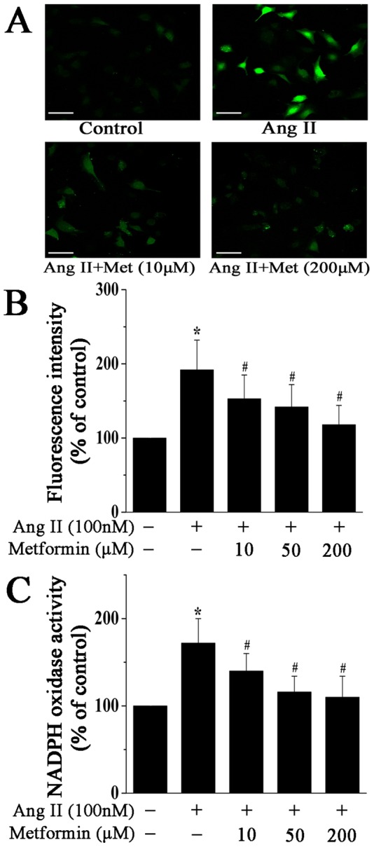
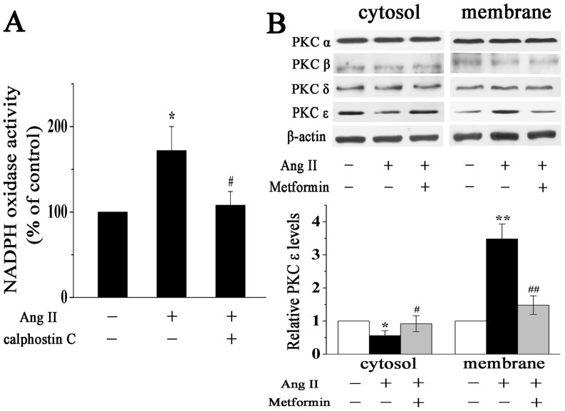
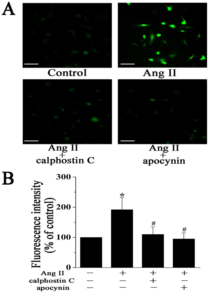
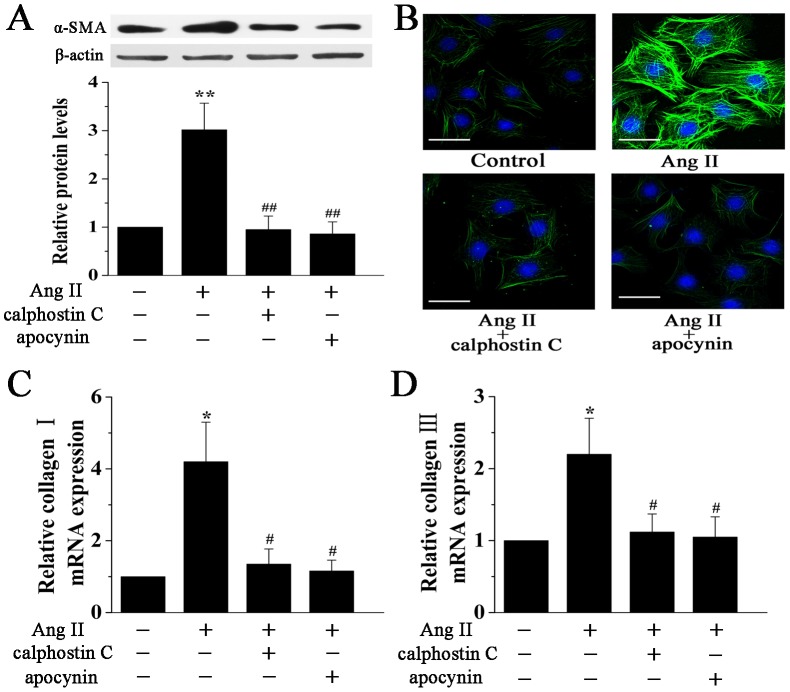
Differentiation of cardiac fibroblasts into myofibroblasts is a critical event in the progression of cardiac fibrosis that leads to pathological cardiac remodeling. Metformin, an antidiabetic agent, exhibits a number of cardioprotective properties. However, much less is known regarding the effect of metformin on cardiac fibroblast differentiation. Thus, in the present study, we examined the effect of metformin on angiotensin (Ang) II-induced differentiation of cardiac fibroblasts into myofibroblasts and its underlying mechanism. Adult rat cardiac fibroblasts were stimulated with Ang II (100 nM) in the presence or absence of metformin (10-200 µM). Ang II stimulation induced the differentiation of cardiac fibroblasts into myofibroblasts, as indicated by increased expression of α-smooth muscle actin (α-SMA) and collagen types I and III, and this effect of Ang II was inhibited by pretreatment of cardiac fibroblasts with metformin. Metformin also decreased Ang II-induced reactive oxygen species (ROS) generation in cardiac fibroblasts via inhibiting the activation of the PKC-NADPH oxidase pathway. Further experiments using PKC inhibitor calphostin C and NADPH oxidase inhibitor apocynin confirmed that inhibition of the PKC-NADPH oxidase pathway markedly attenuated Ang II-induced ROS generation and myofibroblast differentiation. These data indicate that metformin inhibits Ang II-induced myofibroblast differentiation by suppressing ROS generation via the inhibition of the PKC-NADPH oxidase pathway in adult rat cardiac fibroblasts. Our results provide new mechanistic insights regarding the cardioprotective effects of metformin and provide an efficient therapeutic strategy to attenuate cardiac fibrosis.


Conflict of interest statement

Competing Interests: The authors have declared that no competing interests exist.

Figures

Figure 1
Figure 1. Metformin inhibits Ang II-induced cardiac myofibroblast differentiation.
A, Representative Western blotting (upper panel) and quantitative analysis (lower panel) of α-SMA protein expression in cardiac fibroblasts treated with Ang II (100 nM) for 24 h in the presence or absence of metformin (10, 50, and 200 µM). α-SMA expression was normalized to β-actin. B, Representative images of immunofluorescence staining for α-SMA (green) in cardiac fibroblasts treated as above. Nuclei were stained with DAPI (blue). Scale bar, 50 µm. C and D, Real-time PCR quantification of Collagen I (A) and Collagen III (B) expression in cardiac fibroblasts treated with Ang II (100 nM) for 12 h in the presence or absence of metformin (10, 50, and 200 µM). The mRNA expression was normalized to corresponding β-actin mRNA. Data represent mean ± SEM of 4 separate experiments. *P<0.05 vs control group, #P<0.05 vs Ang II group, **P<0.01 vs control group, ##P<0.01 vs Ang II group.
Figure 2
Figure 2. Metformin inhibits Ang II-induced ROS generation and NADPH oxidase activity in cardiac fibroblasts.
A, Representative images of DCF fluorescence (green) in cardiac fibroblasts treated with Ang II (100 nM) for 12 h in the presence or absence of metformin (10 and 200 µM). Scale bar, 100 µm. B, Quantitative analysis of DCF fluorescence intensity by flow cytometry. C, Quantitative analysis of NADPH oxidase activity in cardiac fibroblasts treated as above. Data represent mean ± SEM of 5 separate experiments. *P<0.05 vs control group, #P<0.05 vs Ang II group.
Figure 3
Figure 3. Metformin prevents Ang II-induced activation of PKC in cardiac fibroblasts.
A, PKC mediates Ang II-induced activation of NADPH oxidase. Quantitative analysis of NADPH oxidase activity in cardiac fibroblasts treated with Ang II (100 nM) for 12 h in the presence or absence of PKC inhibitor calphostin C. B, Representative Western blotting (upper panel) and quantitative analysis (lower panel) of PKC isoforms expression in the cytosolic and membrane fractions of cardiac fibroblasts treated with Ang II (100 nM) for 12 h in the presence or absence of metformin (200 µM). PKC ε expression was normalized to β-actin. Data represent mean ± SEM of 5 separate experiments. *P<0.05 vs control group, #P<0.05 vs Ang II group, **P<0.01 vs control group, ##P<0.01 vs Ang II group.
Figure 4
Figure 4. The PKC-NADPH oxidase pathway mediates Ang II-induced ROS generation in cardiac fibroblasts.
A, Representative images of DCF fluorescence (green) in cardiac fibroblasts treated with Ang II (100 nM) for 12 h in the presence or absence of 0.1 µM calphostin C (PKC inhibitor) or 1 mM apocynin (NADPH oxidase inhibitor). Scale bar, 100 µm. B, Quantitative analysis of DCF fluorescence intensity by flow cytometry. Data represent mean ± SEM of 5 separate experiments. *P<0.05 vs control group, #P<0.05 vs Ang II group.
Figure 5
Figure 5. The PKC-NADPH oxidase pathway mediates Ang II-induced cardiac myofibroblast differentiation.
A, Representative Western blotting (upper panel) and quantitative analysis (lower panel) of α-SMA protein expression in cardiac fibroblasts treated with Ang II (100 nM) for 24 h in the presence or absence of calphostin C (0.1 µM) or apocynin (1 mM). α-SMA expression was normalized to β-actin. B, Representative images of immunofluorescence staining for α-SMA (green) in cardiac fibroblasts treated as above. Nuclei were stained with DAPI (blue). Scale bar, 50 µm. C and D, Real-time PCR quantification of Collagen I (A) and Collagen III (B) expression in cardiac fibroblasts treated with Ang II (100 nM) for 12 h in the presence or absence of calphostin C (0.1 µM) or apocynin (1 mM). The mRNA expression was normalized to corresponding β-actin mRNA. Data represent mean ± SEM of 4 separate experiments. *P<0.05 vs control group, #P<0.05 vs Ang II group, **P<0.01 vs control group, ##P<0.01 vs Ang II group.

References

    1. Porter KE, Turner NA (2009) Cardiac fibroblasts: at the heart of myocardial remodeling. Pharmacol Ther 123: 255–278. - PubMed
    1. Vasquez C, Benamer N, Morley GE (2011) The cardiac fibroblast: functional and electrophysiological considerations in healthy and diseased hearts. J Cardiovasc Pharmacol 57: 380–388. - PMC - PubMed
    1. Weber KT (2004) Fibrosis in hypertensive heart disease: focus on cardiac fibroblasts. J Hypertens 22: 47–50. - PubMed
    1. Petrov VV, Fagard RH, Lijnen PJ (2002) Stimulation of collagen production by transforming growth factor-beta1 during differentiation of cardiac fibroblasts to myofibroblasts. Hypertension 39: 258–263. - PubMed
    1. Holtz J (1993) Pathophysiology of heart failure and the renin-angiotensin-system. Basic Res Cardiol 88 Suppl 1183–201. - PubMed

Publication types

LinkOut - more resources