. 2013 Sep 4;8(9):e73260.
doi: 10.1371/journal.pone.0073260. eCollection 2013.

Generation and characterization of a diabody targeting the αvβ6 integrin

Affiliations

Generation and characterization of a diabody targeting the αvβ6 integrin

Heide Kogelberg et al. PLoS One. .

Abstract

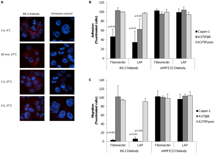
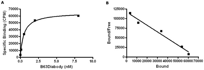
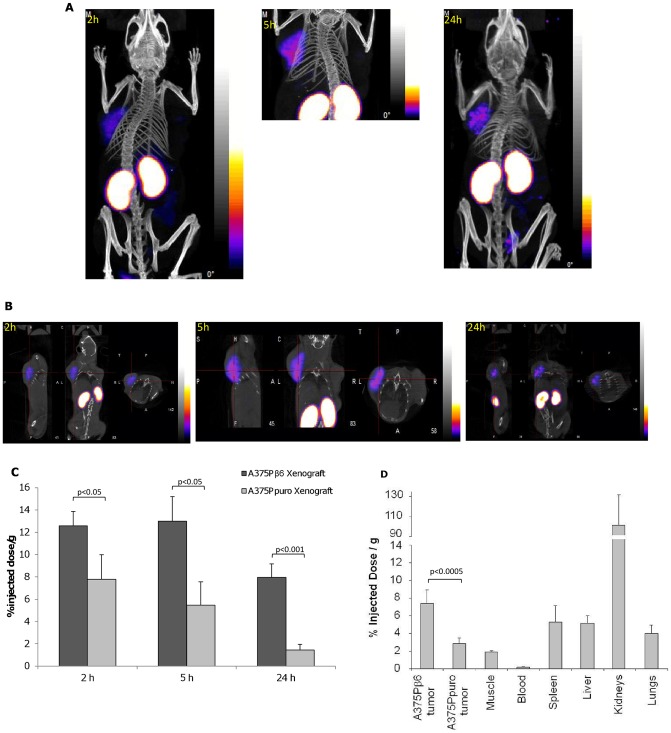
The αvβ6 integrin is up-regulated in cancer and wound healing but it is not generally expressed in healthy adult tissue. There is increasing evidence that it has a role in cancer progression and will be a useful target for antibody-directed cancer therapies. We report a novel recombinant diabody antibody fragment that targets specifically αvβ6 and blocks its function. The diabody was engineered with a C-terminal hexahistidine tag (His tag), expressed in Pichia pastoris and purified by IMAC. Surface plasmon resonance (SPR) analysis of the purified diabody showed affinity in the nanomolar range. Pre-treatment of αvβ6-expressing cells with the diabody resulted in a reduction of cell migration and adhesion to LAP, demonstrating biological function-blocking activity. After radio-labeling, using the His-tag for site-specific attachment of (99m)Tc, the diabody retained affinity and targeted specifically to αvβ6-expressing tumors in mice bearing isogenic αvβ6 +/- xenografts. Furthermore, the diabody was specifically internalized into αvβ6-expressing cells, indicating warhead targeting potential. Our results indicate that the new αvβ6 diabody has a range of potential applications in imaging, function blocking or targeted delivery/internalization of therapeutic agents.


Conflict of interest statement

Competing Interests: The authors have declared that no competing interests exist.

Figures

Figure 1
Figure 1. Production of B6.3 diabody and analysis of its specific interaction with αvβ6.
A) Size-exclusion chromatographic profile (Superdex 75, 125 ml) of B6.3 diabody after fermentation, expanded-bed adsorption IMAC, Superdex 75 (500 ml), 1 ml Ni2+-charged Hi-Trap IMAC, freezing and de-frosting. B6.3 diabody eluted from the column as a dimer that separated in monomeric form under reducing conditions by SDS-PAGE, consistent with non-covalent association of monomers in a diabody structure. B) Sensogram of real-time binding and dissociation of B6.3 diabody to αvβ6. B6.3 diabody was immobilized on a BIAcore CM5 sensor chip and αvβ6 protein was flown across at 400, 200, 100, 50, 25, 12.5, 6.25 and 3.125 nM. The affinity constant (KD) for the interaction was 2.8×10⁻⁹ M, with on-rate of 8,107±7.3 M−1s−1 and off-rate of 2.3×10⁻⁵±1.4×10⁻⁷ s−1. C) Flow cytometry analysis of B6.3 diabody binding to αvβ6-expressing A375Pβ6 cells in a concentration-dependent manner (a) but not to A375Ppuro cells (b), which do not express this integrin. Cells were incubated with B6.3 diabody, at the indicated concentrations and binding was detected with mouse anti-tetra-histidine IgG followed by R-PE-labeled goat anti-mouse IgG. B6.3 diabody was not added to omission control (shown in solid grey). D) Inhibition of B6.3 diabody binding after incubation with the anti-αvβ6 antibody 10D5 shown by flow cytometry. Cells were incubated with 100 ng B6.3 diabody with or without prior incubation with 10D5 at the indicated concentrations. Binding of B6.3 diabody was detected with rabbit anti-hexahistidine IgG followed by R-PE-labeled goat anti-rabbit IgG. In the omission control experiment (shown in solid grey) cells were not incubated with B6.3 diabody and 10D5.
Figure 2
Figure 2. Treatment of αvβ6-expressing cells with B6.3 diabody resulted in diabody internalization and blockade of integrin functions.
A) Localization of B6.3 diabody in A375Pβ6 cells by confocal microscopy. B6.3 diabody detection showed membrane pattern of staining at 4°C and internalized when cells were incubated at 37°C for 30 min, 1 h and 3 h. B6.3 diabody was detected using rabbit anti-human IgG followed by Alexa Fluor 546®-labeled goat anti-rabbit IgG (red). Cells were also counterstained with Hoechst 33245 (blue). B) Treatment of αvβ6-expressing cells blocked adhesion to LAP-coated plates (A375Pβ6 and Capan-1 cells) and/or fibronectin-coated plates (Capan-1 cells). Cells were incubated with B6.3 or shMFE23 diabody at 4°C for 1 h and allowed to attach to coated plates for 1 h at 37°C. Treatment with the anti-CEA shMFE23 diabody had no effect on the cell lines used. C) B6.3 diabody treatment inhibited migration towards LAP and fibronectin. As observed in adhesion assays, the diabody inhibited migration of A375Pβ6 cells to LAP and migration of Capan-1 cells to fibronectin and LAP, while targeting CEA had no effect on the cells tested.
Figure 3
Figure 3. B6.3 diabody inhibited LAP-mediated Smad2/3 translocation to the nucleus in Capan-1 cells.
Cells were incubated at 4°C in the presence of B6.3 diabody and then treated with LAP or TGFβ1 (30 min, 37°C); Smad2/3 localization was assessed by confocal microscopy (40X) using rabbit anti-Smad2/3 followed by Alexa Fluor 488®-labeled goat anti-rabbit IgG (green); smad2/3 was found in the cytoplasm of starved Capan-1 cells (a) and after treatment with B6.3 diabody (c). Smad2/3 was present in the nuclei in response to treatment with latent TGFβ1 (b), a translocation that was inhibited by pre-treatment B6.3 diabody (d). TGFβ1 was used as a positive control (e.f).
Figure 4
Figure 4. Labeling with 99mTc did not affect B6.3 diabody binding to αvβ6.
A) Saturation Binding experiment showed concentration-dependent binding of 99mTc-labeled diabody to A375Pβ6 cells. Non-specific binding, including 25 µg of unlabeled diabody was subtracted from each data point. KD obtained was 4.88±0.32×10⁻⁸ M and BMax was 2.3±0.039×10⁵ receptors/cell (325±5.53 pM/8.5×105 cells). B) Scatchard presentation of the data. Each experiment was carried out in duplicate.
Figure 5
Figure 5. 99mTc-labeled B6.3 diabody localised specifically to αvβ6-expressing tumors in vivo.
A) A375Pβ6 and A375Ppuro cells were injected subcutaneously on opposite shoulders and 99mTc-labeled B6.3 diabody (approximately 11 µg, 30 MBq) was injected intravenously once tumours had developed. Mice were imaged by SPECT/CT as indicated 2 h, 5 h and 24 h after injection. B) SPECT/CT cross sections of the same mice at 2, 5 and 24 h. C) Percent injected doses of 99mTc-labeled B6.3 diabody in A375Pβ6 and A375Ppuro tumours from three mice, obtained from these images. D) Biodistribution of 99mTc-labeled B6.3 diabody 24 h after injection. Data expressed as % injected dose/g (%ID/g) as mean ± SD for 5 animals. Tumor-to blood ratios at this time point were 40.4 for A375Pβ6 tumors and 15.5 for A375Ppuro tumors. Significance assessed by Student's t-test.

References

    1. Patsenker E, Wilkens L, Banz V, Osterreicher CH, Weimann R, et al. (2010) The alphavbeta6 integrin is a highly specific immunohistochemical marker for cholangiocarcinoma. J Hepatol 52: 362–369. - PubMed
    1. Bandyopadhyay A, Raghavan S (2009) Defining the role of integrin alphavbeta6 in cancer. Curr Drug Targets 10: 645–652. - PMC - PubMed
    1. Sipos B, Hahn D, Carceller A, Piulats J, Hedderich J, et al. (2004) Immunohistochemical screening for beta6-integrin subunit expression in adenocarcinomas using a novel monoclonal antibody reveals strong up-regulation in pancreatic ductal adenocarcinomas in vivo and in vitro. Histopathology 45: 226–236. - PubMed
    1. Bates RC (2005) Colorectal cancer progression: integrin alphavbeta6 and the epithelial-mesenchymal transition (EMT). Cell Cycle 4: 1350–1352. - PubMed
    1. Hazelbag S, Kenter GG, Gorter A, Dreef EJ, Koopman LA, et al. (2007) Overexpression of the alpha v beta 6 integrin in cervical squamous cell carcinoma is a prognostic factor for decreased survival. J Pathol 212: 316–324. - PubMed

Publication types

MeSH terms