. 2013 Sep 2;8(9):e73262.
doi: 10.1371/journal.pone.0073262. eCollection 2013.

Expression of Livin in colorectal cancer and its relationship to tumor cell behavior and prognosis

Affiliations

Expression of Livin in colorectal cancer and its relationship to tumor cell behavior and prognosis

Dae-Seong Myung et al. PLoS One. .

Abstract

Backgrounds: Expression of Livin, a member of the inhibitors of apoptosis protein family, is associated with tumor development and progression. The aims of this study were to evaluate whether Livin affects oncogenic biological behavior of colorectal cancer cells, and to document the relationship between its expression and various clinicopathological parameters in colorectal cancer.

Methods: We investigated the impact of Livin on tumor cell behavior by using the small interfering RNA and pcDNA3.1 vector in SW480 and DKO1 colorectal cancer cell lines. The expression of Livin was investigated by RT-PCR and immunohistochemistry in coloretcal cancer tissues. The apoptotic cells were visualized by TUNEL assay, and proliferative cells were visualized by Ki-67 antibody staining.

Results: Knockdown of Livin suppressed tumor cell migration and invasion in colorectal cancer cells. Knockdown of Livin induced the apoptosis by up-regulating of caspase-3, -7 and PARP activities and the cell cycle arrest by decreasing cyclin D1, cyclin D3, cyclin-dependent kinase 4 and 6, and by inducing p27 expression. The MAPK signaling cascades were significantly blocked by knockdown of Livin. In contrast, overexpression of Livin enhanced tumor cell migration and invasion, and inhibited the apoptosis and cell cycle arrest. The mean apoptotic index (AI) value of Livin positive tumors was significantly lower than AI of Livin negative tumors. However, there was no significant difference between Livin expression and Ki-67 labeling index (KI). Livin expression was significantly increased in colorectal cancer and metastatic lymph node tissues compared to normal colorectal mucosa and non-metastatic lymph node tissues and was associated with tumor stage, lymphovascular invasion, lymph node metastasis and poor survival.

Conclusions: These results indicate that Livin is associated with tumor progression by increasing tumor cell motility and inhibiting apoptosis in colorectal cancer.


Conflict of interest statement

Competing Interests: The authors have declared that no competing interests exist.

Figures

Figure 1
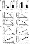
Figure 1. The impact of Livin on invasion, migration and proliferation of human colorectal cancer cells.
(A) The impact of Livin on invasion of colorectal cancer cells. The invasion assay using the siRNA or pcDNA3.1-transfected cells was performed. Stained invading cells were counted and are represented as a graph between groups. The number of LS-transfected cells that invaded was significantly lower than that of SS-transfected cells. The number of invading cells was significantly higher in LV-transfected cells compared to EV-transfected cells (mean±standard error [SE], n=6; *p<0.05). (B) The impact of Livin on migration of colorectal cancer cells. The wound healing assay using siRNA or pcDNA3.1-transfected cells was performed and graphs of cell migration are displayed as relative healing distances. Cell migration was significantly disturbed in LS-transfected SW480 and DKO1 cells and increased in LV-transfected SW480 cells (mean±SE, n=3; *p<0.05). (C) The impact of the Livin on proliferation of colorectal cancer cells. The absorbance indicating proliferating viable cells decreased in the LS-transfected SW480 cells but there was no significant difference of cell proliferation between LV and EV transfected cells (mean±SE, n=3; *p<0.05). SS; scramble siRNA, LS; Livin siRNA, EV; Empty-pcDNA3.1, LV; pcDNA3.1-Livin.
Figure 2
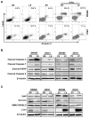
Figure 2. The impact of Livin on apoptosis in human colorectal cancer cells.
(A) The proportion of apoptotic cells induced by transfection of LS was greater than that induced by transfection of SS (6.9 vs. 19.6%) in SW480 cells but Livin knockdown had a minimal influence on apoptosis (11.3 vs. 16.7%) in DKO1 cells. Overexpression of Livin by transfection of LV inhibited the apoptosis of SW480 cells in response to 5-FU but overexpression of Livin had a minimal influence on apoptosis in DKO1 cells (B) Expression of cleaved caspase-3, -7, -9, and PARP proteins. The caspase-3, -7 and PARP expression was increased in SW480 and DKO1 cells after Livin knockdown, and decreased after overexpression of Livin (C) Expression of apoptosis regulatory proteins. Survivin protein level decreased following Livin knockdown in SW480 and DKO1 cells, but XIAP and SMAC/DIABLO protein levels were not altered in response to Livin knockdown. Additionally, Survivin, XIAP and SMAC/DIABLO protein levels were not altered after overexpression of Livin. PARP; Poly (ADP-ribose) polymerase, XIAP; X-chromosome binding IAP, SMAC/DIABLO; second mitochondria-derived activator of caspases/direct IAP binding protein with low pI, SS; scramble siRNA, LS; Livin siRNA, EV; Empty-pcDNA3.1, LV; pcDNA3.1-Livin, 5-FU; 5-fluorouracil, 7-AAD; 7-amino-actinomycin D.
Figure 3
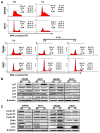
Figure 3. The impact of Livin on cell cycle arrest in human colorectal cancer cells.
(A) Livin knockdown resulted in cell cycle arrest in the G0/G1 phase of SW480 cells and the S phase of DKO1 cells. 5-FU treatment induced cell cycle arrest in the subG1 phase of SW480 and the G0/G1 phase of DKO1 cells. Overexpression of Livin inhibited 5-FU-induced cell cycle arrest in SW480 cells and had a minimal influence in DKO1 cells. One representative experiment of the three independent experiments is shown. (B) Expression of cyclin-dependent kinase (CDK) inhibitor proteins. The p27 protein level was significantly increased by Livin knockdown and decreased by overexpression of Livin in SW480 and DKO1 cells. However, p21, p57, p15 and p16 protein levels were not altered in response to knockdown or overexpression of Livin. (C) Expression of cyclins and cyclin-dependent kinase (CDKs) proteins. Cyclin D1, cyclin D3, CDK4 and CDK6 protein levels were significantly decreased by Livin knockdown and increased the cyclin D1 and CDK4 by overexpression of Livin in SW480 and DKO1 cells. SS; scramble siRNA, LS; Livin siRNA, EV; Empty-pcDNA3.1, LV; pcDNA3.1-Livin.
Figure 4
Figure 4. The impact of Livin on intracellular signaling pathways involved in apoptosis and cell cycle arrest of human colorectal cancer cells.
The phosphorylation levels of ERK1/2, JNK and p38 decreased following Livin knockdown of SW480 and DKO1 cells. But Akt and p65 the phosphorylation levels showed no change following Livin knockdown. Additionally, ERK1/2, JNK, p38, Akt and p65 phosphorylation levels were not changed by overexpression of Livin. SS; scramble siRNA, LS; Livin siRNA, EV; Empty-pcDNA3.1, LV; pcDNA3.1-Livin.
Figure 5
Figure 5. Expression of Livin mRNA and protein in fresh colonoscopic biopsy specimens.
(A) Livin mRNA expression (B) Livin protein expression. Expression of Livin is upregulated in cancer tissues compared to paired normal mucosa at mRNA and protein levels in fresh colonoscopic biopsy specimens. Each bar represents the mean ± SE of 20 cases. *p<0.05 versus normal colorectal mucosa. T; colorectal cancer tissue, N; paired normal colorectal mucosa.
Figure 6
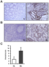
Figure 6. Livin protein expression in human colorectal cancer and metastatic lymph node tissues by immunohistochemistry.
(A) The Livin protein was predominantly immunostained in the nuclei of cancer cells but did not or only weakly immunostained in normal colorectal mucosa (× 100). (B) The immunostaining of Livin in metastatic lymph node tissues was significantly stronger than that in non-metastatic lymph node tissues (× 100). (C) The overall score for Livin immunostaining in metastatic lymph node tissues was significantly higher than that in non-metastatic lymph node tissues (*p<0.001). T; colorectal cancer tissue, N; paired normal colorectal mucosa, NL; non-metastatic lymph node tissue, ML; metastatic lymph node tissue.
Figure 7
Figure 7. Assessment of tumor cell proliferation and apoptosis in human colorectal cancers.
(A) Immunostaining of Ki-67. Immunostaining of Ki-67 shows strong nuclear positivity in the cancer cells but is rarely positive in the normal colorectal mucosa (× 200). (B) Detection of apoptotic cells (arrow) by TUNEL staining. Apoptotic cells with classic features of DNA condensation are shown to have a halo consisting of pyknotic nucleus and shrunken cytoplasm (arrow head) in colorectal cancer tissues but TUNEL staining is not positive in normal colorectal mucosa (× 400). TUNEL, terminal deoxynucleotidyl transferase (TdT)-mediated deoxyuridine triphosphate (dUTP) nick end labeling. T; colorectal cancer tissue, N; paired normal colorectal mucosa.
Figure 8
Figure 8. Kaplan-Meier survival curve correlating overall survival with positive expression (solid line) and negative expression (dotted line) of Livin (p=0.014).

References

    1. Labianca R, Merelli B (2010) Screening and diagnosis for colorectal cancer: present and future. Tumori 96: 889-901. PubMed: 21388049. - PubMed
    1. Ahmed FE (2005) Development of novel diagnostic and prognostic molecular markers for sporadic colon cancer. Expert Rev Mol Diagn 5: 337-352. doi:10.1586/14737159.5.3.337. PubMed: 15934812. - DOI - PubMed
    1. Allen WL, Johnston PG (2005) Have we made progress in pharmacogenomics? The implementation of molecular markers in colon cancer. Pharmacogenomics 6: 603-614. doi:10.2217/14622416.6.6.603. PubMed: 16143000. - DOI - PubMed
    1. Riethdorf S, Wikman H, Pantel K (2008) Review: Biological relevance of disseminated tumor cells in cancer patients. Int J Cancer 123: 1991-2006. doi:10.1002/ijc.23825. PubMed: 18712708. - DOI - PubMed
    1. Jankowski JA, Odze RD (2009) Biomarkers in gastroenterology: between hope and hype comes histopathology. Am J Gastroenterol 104: 1093-1096. doi:10.1038/ajg.2008.172. PubMed: 19417749. - DOI - PubMed

Publication types

MeSH terms

Substances