. 2013 Sep 4;8(9):e73752.
doi: 10.1371/journal.pone.0073752. eCollection 2013.

DEAD box protein DDX1 regulates cytoplasmic localization of KSRP

Affiliations

DEAD box protein DDX1 regulates cytoplasmic localization of KSRP

Chu-Fang Chou et al. PLoS One. .

Abstract

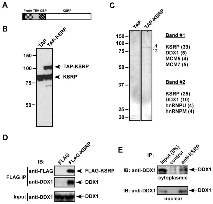
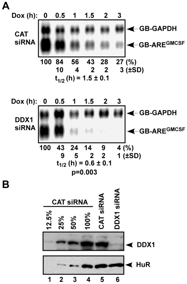
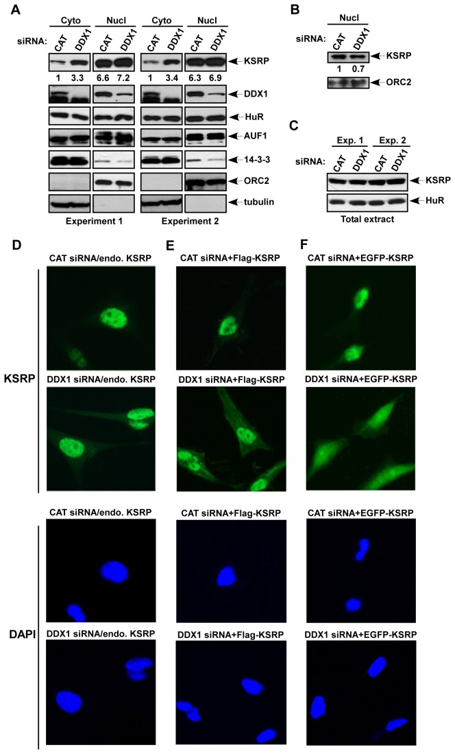
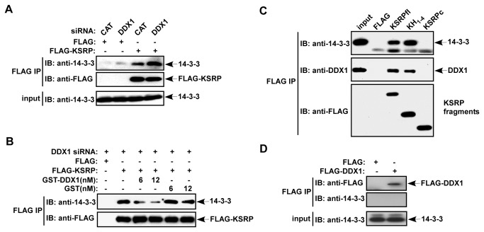
mRNA decay mediated by the AU-rich elements (AREs) is one of the most studied post-transcriptional mechanisms and is modulated by ARE-binding proteins (ARE-BPs). To understand the regulation of K homology splicing regulatory protein (KSRP), a decay-promoting ARE-BP, we purified KSRP protein complexes and identified an RNA helicase, DDX1. We showed that down-regulation of DDX1 expression elevated cytoplasmic levels of KSRP and facilitated ARE-mediated mRNA decay. Association of KSRP with 14-3-3 proteins, that are predominately located in the cytoplasm, increased upon reduction of DDX1. We also demonstrated that KSRP associated with DDX1 or 14-3-3, but not both. These observations indicate that subcellular localization of KSRP is regulated by competing interactions with DDX1 or 14-3-3.


Conflict of interest statement

Competing Interests: The authors have declared that no competing interests exist.

Figures

Figure 1
Figure 1. Purification of KSRP-associated proteins.
(A) A schematic diagram of TAP-KSRP. Sequences encoding the TAP tag containing protein A (ProtA), a TEV protease cleavage site, and a calmodulin binding peptide (CBP) were fused to the N-terminus of KSRP. (B) Extracts from HT1080 stable cells expressing the TAP tag or TAP-KSRP were analyzed by anti-KSRP immunoblotting. (C) Extracts containing TAP and TAP-KSRP were treated with RNase A and subjected to TAP purification. The purified fractions were analyzed by silver staining. Two bands detected only in the TAP-KSRP fraction are labeled (1 and 2). Proteins identified from bands 1 and 2 by mass spectrometry are indicated. Numbers of observed peptides are denoted in parentheses. (D) FLAG or FLAG-KSRP was expressed in HeLa-TO cells. RNase A-treated extracts were subjected to anti-FLAG immunoprecipitation. The precipitates were analyzed by anti-FLAG or anti-DDX1 immunoblotting. 5% of input used for immunoprecipitation was also analyzed by anti-DDX1. (E) RNAse A-treated cytoplasmic and nuclear extracts were immunoprecipitated with an anti-KSRP monoclonal antibody or a control IgG. The precipitates and 5% of input were analyzed by anti-DDX1 immunoblotting.
Figure 2
Figure 2. Down-regulation of DDX1 facilitates AMD.
(A) HeLa-TO cells were transfected with a construct expressing GB-AREGMCSF mRNA under the control of a Tet-regulatory promoter and a construct constitutively expressing GB-GAPDH mRNA under the control of the CMV promoter. The cultures were also transfected a control siRNA (CAT) or a DDX1 siRNA. Cytoplasmic RNA was isolated at different time points after the addition of doxycycline (Dox). The levels of GB-AREGMCSF and GB-GAPDH mRNAs were analyzed by Northern blot. Signals of GB-AREGMCSF mRNA were quantified by a phosphorimager and normalized to that of GB-GAPDH mRNA. The calculated half-lives (t1/2; n=3) of GB-AREGMCSF mRNA are shown as mean values ± standard deviations from three independent experiments. P value is indicated and calculated by Student’s t-test using Microsoft Excel software. (B) Downregulation of DDX1 by siRNA. Extracts of HeLa-TO cells in (A) were subjected to immunoblot analysis with anti-DDX1 or anti-HuR antibodies. Different amounts of CAT siRNA-treated extracts (12, 25, 50, or 100% of the amounts used in lane 5) were loaded to estimate knockdown efficiency.
Figure 3
Figure 3. Subcellular localization of KSRP is regulated by DDX1.
(A) HeLa-TO cells were transfected with a control siRNA or a DDX1 siRNA. Cytoplasmic and nuclear extracts from equal number of cells were subjected to immunoblot analysis with anti-KSRP, anti-DDX1, anti-HuR, anti-AUF1, or anti-14-3-3 which recognizes all isoforms. Antibodies against cytoplasmic α-tubulin and a nuclear protein, origin recognition complex subunit 2 (ORC2), were also used as controls for subcellular fractionation. Two independent transfection experiments were carried out. Quantification of KSRP levels in the cytoplasmic and nuclear fractions is indicated. (B) The nuclear extracts used in (A) were diluted 5-fold and subjected to immunoblot analysis with anti-KSRP and anti-ORC2. Quantification of the nuclear KSRP levels is indicated. (C) Total extracts of cells transfected with CAT or DDX1 siRNAs were analyzed by anti-KSRP or anti-HuR (D to F). HeLa-TO cells were transfected with CAT siRNA or DDX1 siRNA (D), CAT siRNA or DDX1 siRNA and a construct expressing FLAG-KSRP (E), or CAT siRNA or DDX1 siRNA and a construct expressing EGFP-KSRP (F). Transfected cells were analyzed by anti-KSRP (D), anti-FLAG (E), or GFP signal (F) (top two rows), and by DAPI staining (bottom two rows).
Figure 4
Figure 4. DDX1 interferes with KSRP association with 14-3-3 and competes with 14-3-3 for interaction with KSRP.
(A) HeLa-TO cells were cotransfected with a FLAG-KSRP expression vector or a control FLAG vector and a control siRNA or a DDX1 siRNA. Cytoplasmic extracts were subjected to anti-FLAG immunoprecipitation. The precipitates were analyzed by anti-14-3-3 and anti-FLAG immunoblotting. 5% input was also analyzed by anti-14-3-3. (B) HeLa-TO cells were cotransfected with a FLAG-KSRP expression vector or a control FLAG vector and DDX1 siRNA. Cytoplasmic extracts were subjected to anti-FLAG immunoprecipitation with the addition of two different concentrations (6 and 12 nM) of recombinant GST-DDX1 or GST. The precipitates were analyzed by anti-14-3-3 and anti-FLAG immunoblotting. (C) HeLa-TO cells were transfected with vectors expressing FLAG-KSRP or FLAG-KSRP fragments consisting of the four KH motifs or the C-terminus. The FLAG immunoprecipitations were subjected to immunoblotting with anti-14-3-3, anti-DDX1, and anti-FLAG. (D) HeLa-TO cells were transfected with a control FLAG vector or a FLAG-DDX1 expression vector. Cytoplasmic extracts were subjected to anti-FLAG immunoprecipitation. The precipitates were analyzed by anti-14-3-3 and anti-FLAG immunoblotting. 5% input was also analyzed by anti-14-3-3.

References

    1. Wilusz CJ, Wormington M, Peltz SW (2001) The cap-to-tail guide to mRNA turnover. Nat Rev Mol Cell Biol 2: 237-246. doi:10.1038/35067025. PubMed: 11283721. - DOI - PubMed
    1. Wilusz CJ, Wilusz J (2004) Bringing the role of mRNA decay in the control of gene expression into focus. Trends Genet 20: 491-497. doi:10.1016/j.tig.2004.07.011. PubMed: 15363903. - DOI - PubMed
    1. Chen CY, Shyu AB (1995) AU-rich elements: characterization and importance in mRNA degradation. Trends Biochem Sci 20: 465-470. doi:10.1016/S0968-0004(00)89102-1. PubMed: 8578590. - DOI - PubMed
    1. Bakheet T, Williams BR, Khabar KS (2003) ARED 2.0: an update of AU-rich element mRNA database. Nucleic Acids Res 31: 421-423. doi:10.1093/nar/gkg023. PubMed: 12520039. - DOI - PMC - PubMed
    1. Bakheet T, Williams BR, Khabar KS (2006) ARED 3.0: the large and diverse AU-rich transcriptome. Nucleic Acids Res 34: D111-D114. doi:10.1093/nar/gkj052. PubMed: 16381826. - DOI - PMC - PubMed

Publication types

LinkOut - more resources