

Altered endothelin receptor expression and affinity in spontaneously hypertensive rat cerebral and coronary arteries
- PMID: 24023902
- PMCID: PMC3759417
- DOI: 10.1371/journal.pone.0073761
Altered endothelin receptor expression and affinity in spontaneously hypertensive rat cerebral and coronary arteries
Abstract
Background: Hypertension is associated with arterial hyperreactivity, and endothelin (ET) receptors are involved in vascular pathogenesis. The present study was performed to examine the hypothesis that ET receptors were altered in cerebral and coronary arteries of spontaneously hypertensive rats (SHR).
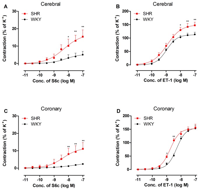
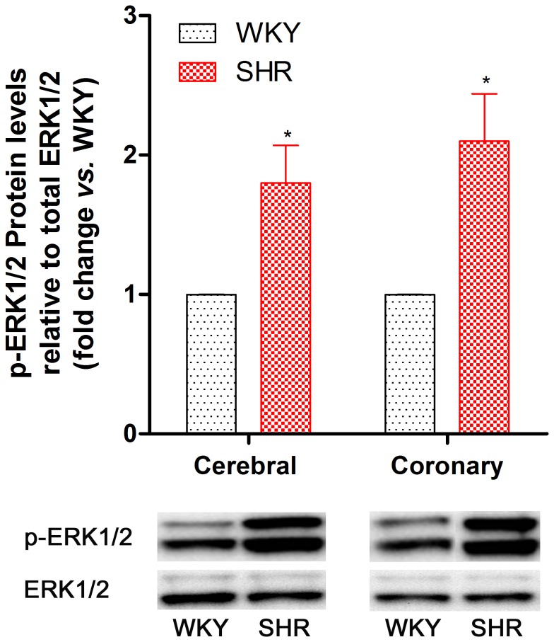
Methodology/principal findings: Cerebral and coronary arteries were removed from SHR. Vascular contraction was recorded using a sensitive myograph system. Real-time PCR and Western blotting were used to quantify mRNA and protein expression of receptors and essential MAPK pathway molecules. The results demonstrated that both ETA and ETB receptor-mediated contractile responses in SHR cerebral arteries were shifted to the left in a nonparallel manner with increased maximum contraction compared with Wistar-Kyoto (WKY) rats. In SHR coronary arteries, the ETA receptor-mediated contraction curve was shifted to the left in parallel with an increased pEC50 compared with the arteries in WKY rats. There was no significant increase in ETB receptor-mediated contraction in SHR coronary arteries. ETA receptor mRNA and protein expression was increased in SHR cerebral arteries compared with the arteries in WKY rats. However, ETA receptor mRNA and protein levels in coronary arteries and ETB receptor protein levels in cerebral and coronary arteries remained unchanged in SHR compared with WKY rats. Meanwhile, phosphorylated ERK1/2 protein was significantly increased in SHR brain and heart vessels.
Conclusions/significance: In SHR cerebral arteries, ETA receptor expression was upregulated. ETA receptor affinity was increased in coronary arteries, and ETB receptor affinity was increased in cerebral arteries. The ERK1/2 activation may be involved in the receptor alterations.
Conflict of interest statement
Figures


References
-
- Zeng C, Villar VA, Eisner GM, Williams SM, Felder RA et al. (2008) G protein-coupled receptor kinase 4: role in blood pressure regulation. Hypertension 51: 1449-1455. doi:10.1161/HYPERTENSIONAHA.107.096487. PubMed: 18347232. - DOI - PMC - PubMed
-
- Edvinsson LI, Povlsen GK (2011) Vascular plasticity in cerebrovascular disorders. J Cereb Blood Flow Metab 31: 1554-1571. doi:10.1038/jcbfm.2011.70. PubMed: 21559027. - DOI - PMC - PubMed
-
- Cline DM (2008) Epidemiology of hypertension. Ann Emerg Med 51: S3-S4. doi:10.1016/S0196-0644(08)00358-2. PubMed: 18191291. - DOI - PubMed
-
- Psaty BM, Furberg CD, Kuller LH, Cushman M, Savage PJ et al. (2001) Association between blood pressure level and the risk of myocardial infarction, stroke, and total mortality: the cardiovascular health study. Arch Intern Med 161: 1183-1192. doi:10.1001/archinte.161.9.1183. PubMed: 11343441. - DOI - PubMed
-
- Agapitov AV, Haynes WG (2002) Role of endothelin in cardiovascular disease. J Renin Angiotensin Aldosterone Syst 3: 1-15. doi:10.3317/jraas.2002.001. PubMed: 11984741. - DOI - PubMed
Publication types
MeSH terms
Substances
LinkOut - more resources
Full Text Sources
Other Literature Sources
Miscellaneous
