. 2013 Sep 4;8(9):e74857.
doi: 10.1371/journal.pone.0074857. eCollection 2013.

Stem cell recruitment of newly formed host cells via a successful seduction? Filling the gap between neurogenic niche and injured brain site

Affiliations

Stem cell recruitment of newly formed host cells via a successful seduction? Filling the gap between neurogenic niche and injured brain site

Naoki Tajiri et al. PLoS One. .

Abstract

Here, we report that a unique mechanism of action exerted by stem cells in the repair of the traumatically injured brain involves their ability to harness a biobridge between neurogenic niche and injured brain site. This biobridge, visualized immunohistochemically and laser captured, corresponded to an area between the neurogenic subventricular zone and the injured cortex. That the biobridge expressed high levels of extracellular matrix metalloproteinases characterized initially by a stream of transplanted stem cells, but subsequently contained only few to non-detectable grafts and overgrown by newly formed host cells, implicates a novel property of stem cells. The transplanted stem cells manifest themselves as pathways for trafficking the migration of host neurogenic cells, but once this biobridge is formed between the neurogenic site and the injured brain site, the grafted cells disappear and relinquish their task to the host neurogenic cells. Our findings reveal that long-distance migration of host cells from the neurogenic niche to the injured brain site can be achieved through transplanted stem cells serving as biobridges for initiation of endogenous repair mechanisms. This is the first report of a stem cell-paved "biobridge". Indeed, to date the two major schools of discipline in stem cell repair mechanism primarily support the concept of "cell replacement" and bystander effects of "trophic factor secretion". The present novel observations of a stem cell seducing a host cell to engage in brain repair advances basic science concepts on stem cell biology and extracellular matrix, as well as provokes translational research on propagating this stem cell-paved biobridge beyond cell replacement and trophic factor secretion for the treatment of traumatic brain injury and other neurological disorders.


Conflict of interest statement

Competing Interests: The authors have read the journal's policy and have the following competing interest: CVB has patents and patent applications on stem cells, their therapeutic applications and mechanisms of action. In particular, the patents and patent applications relate to the use of stem cells for treating human disorders, including the present study's target disease indication of traumatic brain injury. EY, MM and CC are employed by the funder of this study, Sanbio Inc. CVB is supported by National Institutes of Health, National Institute of Neurological Disorders and Stroke 1R01NS071956-01, James and Esther King Foundation for Biomedical Research Program, SanBio Inc., Celgene Cellular Therapeutics, KMPHC and NeuralStem Inc. Senior author CVB is a PLOS ONE Editorial Board member. Patents: 1995 Sertoli cells as neurorecovery inducing cells for neurodegenerative disorders. (US patent: 6,036,951) Inventors: Sanberg, P.R., Borlongan, C.V. & Cameron, D.F. 1995 Sertoli cells as transplantation facilitator for cell transplantation. (US patent: 5,942,437) Inventors: Sanberg, P.R., Borlongan, C.V. & Cameron, D.F. 1996 Method and media for enhancing cryopreservation of cells (US patent: 6,037,175) Inventors: Sanberg, P.R., Borlongan, C.V., & Cameron, D.F. 1999 Melatonin as a protective agent against stroke. (US patent: 6,075,045) Inventors: Nishino, H. & Borlongan, C.V. 2001 Method of attenuating cognitive deficits with methanesulfonyl fluoride. (Patent pending) Inventors: Borlongan, C.V. & Moss, D. 2006 Multipotent progenitor cells rescue against stroke. (IP disclosure at MCG) Inventors: Carroll, J.E., Hess, D.C., & Borlongan, C.V. 2009 Human amnion as a source of stem cells for CNS therapy. (US Patent Pending) Inventors: Borlongan, C.V., & Parollini, O. 2009 Delta opioid peptide for stroke therapy. (US Patent Pending) Inventors: Borlongan, C.V., Su, T.-P., Wang, Y. 2010 Methods for enhancing neuroprotection via administration of stem cells and blood brain barrier permeabilizers. US patent: 7,674,457 B2 Inventors: Borlongan, C.V., and Sanberg, P.R. This does not alter the authors' adherence to all the PLOS ONE policies on sharing data and materials.

Figures

Figure 1
Figure 1. Behavioral tests (performed by two investigators blinded to the treatment condition throughout the study) were initially conducted at baseline (i.e., prior to brain insult) and revealed that all adult SD rats included in this study displayed normal behaviors (A, B, and C).
At 7 days after TBI, the same behavioral tests showed that TBI produced significant impairments in motor and neurological tasks. At one month, two months, and three months post-TBI, transplanted animals displayed significantly improved motor and neurological functions compared to traumatically injured animals that received vehicle only. These behavioral improvements were accompanied by reduction in TBI core and peri-injury cell death (D and E) as revealed by H&E staining (a and b correspond to vehicle and transplant respectively at one month post-treatment, while c and d represent vehicle and transplant respectively at three months post-treatment. a-d are at 10X while a’-d’ at 20X magnification). Asterisks (*) indicate significant improvements in behavioral and histological deficits in TBI transplanted cells compared to TBI animals that received vehicle only (p’s < 0.05).
Figure 2
Figure 2. The biobridge between SVZ and impacted cortex consists of highly proliferative, neurally committed, and migratory cells.
At one month post-TBI, confocal microscopy revealed a surge of proliferative Ki67 positive cells and immature neurally nestin labeled cells in the peri-injured cortical areas (A) and subventricular zone (A’), with a stream of migrating cells (DCX) along the corpus callosum (B) in TBI animals that received the stem cell transplants. In contrast those that received vehicle alone displayed limited cell proliferation (C), neural differentiation (C’), and scattered migration in the peri-injured cortical areas (D) and almost absent expression of newly formed cells in the SVZ (C’).
Figure 3
Figure 3. At three months post-TBI, the brains from vehicle-infused animals displayed a disparate pattern of cell fate in that the newly formed Ki67 positive and nestin labeled cells were sequestered within the corpus callosum (A) and the SVZ (B) and only sporadic cells were able to reach the impacted cortex (A’ and B’), with likely resident DCX cells seen around the impacted cortex (C).
In contrast, at three months post-TBI, the brains from transplanted animals exhibited a much more massive cell proliferation and neural differentiation encasing the peri-injured cortical areas accompanied by a solid stream of nestin (D, D’) and DCX labeled cells (E) migrating not just along, but across the corpus callosum from the SVZ to the impacted cortex.
Figure 4
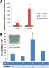
Figure 4. Laser-captured biobridge, corresponding to the brain tissue between SVZ and impacted cortex, expressed high levels of MMP-9 gelatinolytic activities at one month and three months post-TBI in animals transplanted with SB623 which were significantly higher than those TBI animals that received vehicle only or sham-operated animals (*p’s< 0.05 vs. vehicle or sham; Panel A).
Although vehicle-infused TBI animals also showed a significantly upregulated MMP-9 gelatinolytic activity at one month post-TBI (**p< 0.05 vs. sham), the level of this neurovascular proteinase activity reverted back to control-sham levels at three months post-TBI. Each bar represents the mean ± standard deviation from n=3 per treatment group for each time point. Next, to further reveal that SB623 cells promoted cell migration via an ECM-mediated mechanism, primary rat cortical cells were either grown alone or co-cultured with SB623 in the presence or absence of the MMP-9 inhibitor Cyclosporine-A (Panel B). Migratory cell assay (see inset) revealed significantly enhanced migration of primary rat cortical cells into the chamber that contained SB623, which was significantly suppressed by treatment with the inhibitor (*p< 0.05 vs. all other treatment conditions). The absence of SB623 and inhibitor in the cell culture condition, the treatment of the inhibitor alone, and the combined treatment of SB623 and inhibitor did not significantly differ in the resulting cell migratory potential.
Figure 5
Figure 5. After TBI, endogenous repair mechanisms commenced, but are limited to the neurogenic SVZ and to a few quiescent resident neurogenic cells around the impacted cortex (A).
This endogenous repair process is not sufficient to mount a robust and stable defense against the TBI-induced cell death cascade unless exogenous stem cells are introduced. A physical gap between the neurogenic SVZ and the non-neurogenic, impacted cortex prevents migration of neurogenic cells to the injured cortex. Transplantation of stem cells into the peri-injured cortical areas creates a neurovascular matrix of biobridge to bootleg newly formed endogenous cells from the SVZ to the peri-injured cortex (B). Once the biobridge is established, the endogenous repair mechanism is maintained by newly formed host cells even in the absence of stem cells (C). Such transplant-paved biobridge between neurogenic and non-neurogenic sites allows endogenous neurogenic cells to reach injury-specific brain sites.

References

    1. Joyner AL, Skarnes WC, Rossant J (1989) Production of a mutation in mouse En-2 gene by homologous recombination in embryonic stem cells. Nature 338: 153-156. doi:10.1038/338153a0. PubMed: 2563902. - DOI - PubMed
    1. Yasuhara T, Matsukawa N, Hara K, Yu G, Xu L et al. (2006) Transplantation of human neural stem cells exerts neuroprotection in a rat model of Parkinson’s disease. J Neurosci 26: 12497-12511. doi:10.1523/JNEUROSCI.3719-06.2006. PubMed: 17135412. - DOI - PMC - PubMed
    1. Yasuhara T, Hara K, Maki M, Mays RW, Deans RJ et al. (2008) Intravenous grafts recapitulate the neurorestoration afforded by intracerebrally delivered multipotent adult progenitor cells in neonatal hypoxic-ischemic rats. J Cereb Blood Flow Metab 28: 1804-1810. doi:10.1038/jcbfm.2008.68. PubMed: 18594556. - DOI - PMC - PubMed
    1. Borlongan CV, Hadman M, Sanberg CD, Sanberg PR (2004) Central nervous system entry of peripherally injected umbilical cord blood cells is not required for neuroprotection in stroke. Stroke 35: 2385-2389. doi:10.1161/01.STR.0000141680.49960.d7. PubMed: 15345799. - DOI - PubMed
    1. Pastori C, Librizzi L, Breschi GL, Regondi C, Frassoni C et al. (2008) Arterially perfused neurosphere-derived cells distribute outside the ischemic core in a model of transient focal ischemia and reperfusion in vitro. PLOS ONE 3: e2754. doi:10.1371/journal.pone.0002754. PubMed: 18648648. - DOI - PMC - PubMed

Publication types