Review
. 2013 Aug 27;5(8):417-24.
doi: 10.4254/wjh.v5.i8.417.

Imaging appearance of treated hepatocellular carcinoma

Affiliations
Review

Imaging appearance of treated hepatocellular carcinoma

Francesco Agnello et al. World J Hepatol. .

Abstract

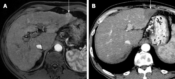
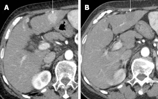
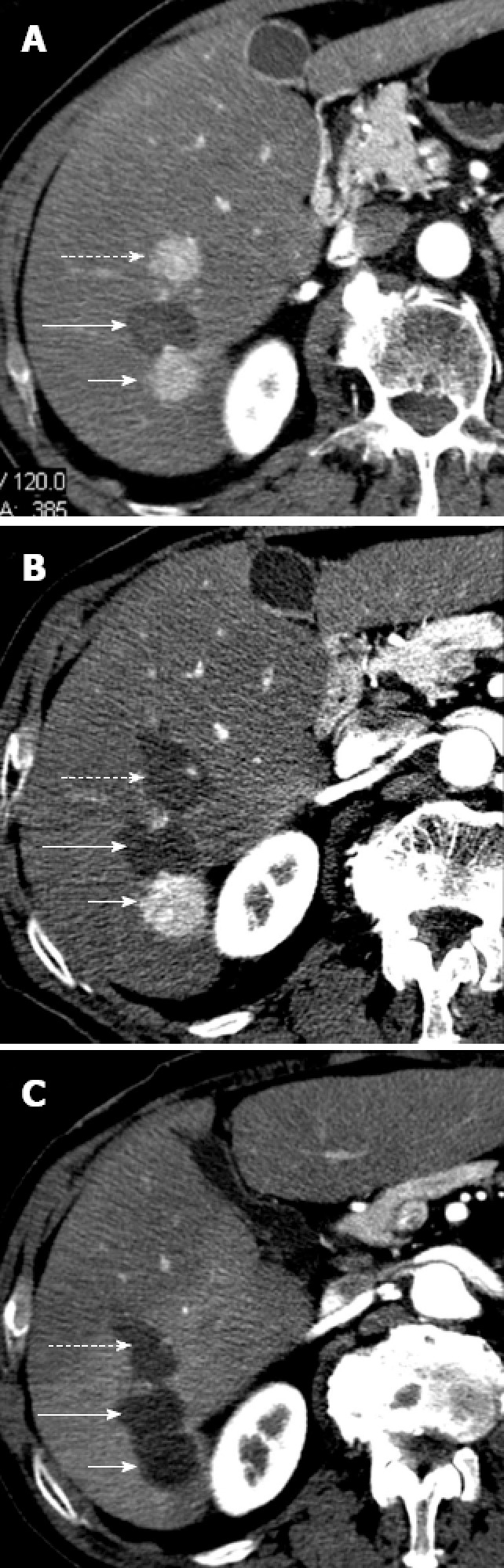
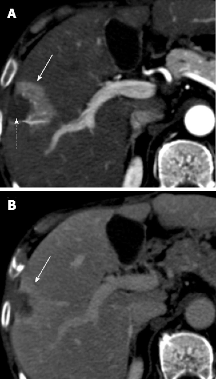
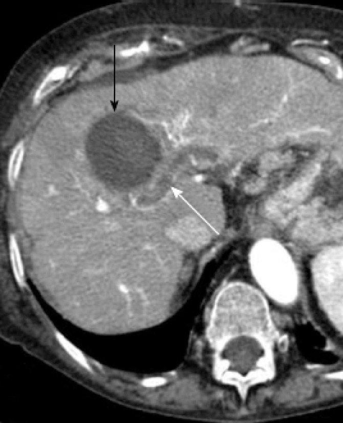
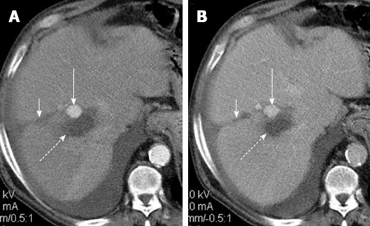
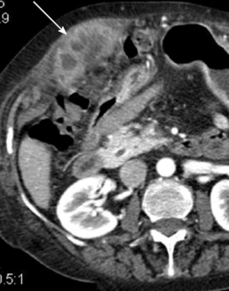
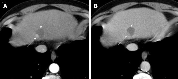
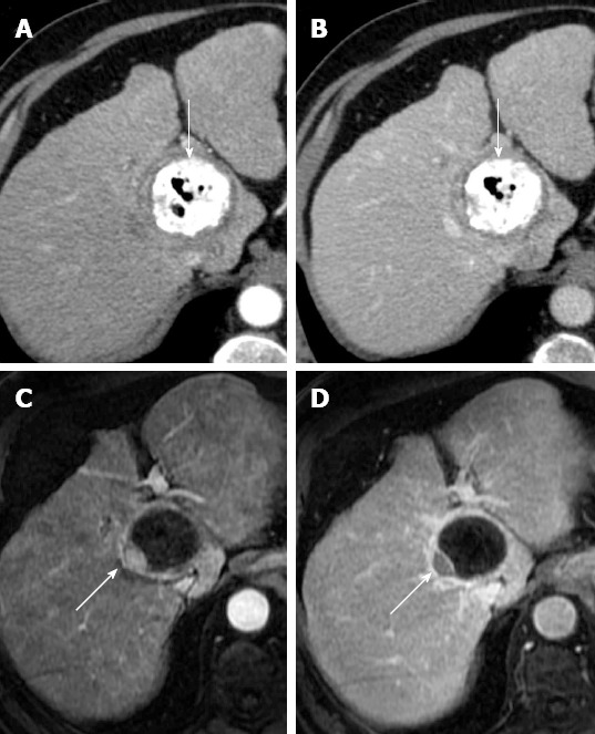
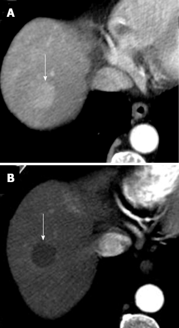
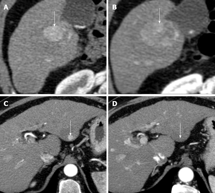
Surgical resection and imaging guided treatments play a crucial role in the management of hepatocellular carcinoma (HCC). Although the primary end point of treatment of HCC is survival, radiological response could be a surrogate end point of survival, and has a key role in HCC decision-making process. However, radiological assessment of HCC treatment efficacy is often controversial. There are few doubts on the evaluation of surgical resection; in fact, all known tumor sites should be removed. However, an unenhancing partial linear peripheral halo, in most cases, surrounding a fluid collection reducing in size during follow-up is demonstrated in successfully resected tumor with bipolar radiofrequency electrosurgical device. Efficacy assessment of locoregional therapies is more controversial and differs between percutaneous ablation (e.g., radiofrequency ablation and percutaneous ethanol injection) and transarterial treatments (e.g., conventional transarterial chemoembolization, transarterial chemoembolization with drug eluting beads and radioembolization). Finally, a different approach should be used for new systemic agent that, though not reducing tumor mass, could have a benefit on survival by delaying tumor progression and death. The purpose of this brief article is to review HCC imaging appearance after treatment.

Keywords: Hepatocellular carcinoma; Imaging; Treatment.


Figures

Figure 1
Figure 1
Successful hepatocellular carcinoma resection with radiofrequency tissue coagulation device. A: Pretreatment arterial phase T1-weighted gradient-echo magnetic resonance shows hypervascular hepatocellular carcinoma (HCC) (arrow); B: On arterial phase computed tomography obtained 4 mo after treatment no hypervascular focus is evident (arrow). Note that resected area is larger to preexisting HCC.
Figure 2
Figure 2
Complete necrosis after radiofrequency ablation for hepatocellular carcinoma. A: Pretreatment arterial phase computed tomography (CT) shows hypervascular hepatocellular carcinoma (arrow); B: Arterial phase CT obtained 1 mo after treatment shows ablated area (arrow). Absence of arterial enhancement suggests complete tumor necrosis.
Figure 3
Figure 3
Hepatocellular carcinoma after multiple radiofrequency ablations. A: Arterial phase computed tomography (CT) obtained 8 mo after radiofrequency ablation shows two new enhancing hepatocellular carcinoma (HCC) nodules located anteriorly (dotted arrow) and posteriorly (short arrow) to ablated HCC (long arrow). These findings suggest occurrence of new HCC nodules; B: Arterial phase CT obtained 2 mo after additional radiofrequency (RF) ablation shows that HCC nodule located anteriorly (dotted arrow) has been replaced by a hypoattenuating, non enhancing area (arrow) that is larger than preexisting tumor. These findings suggest complete necrosis. Posteriorly located HCC (short arrow) increased in size; C: Arterial phase CT obtained 2 mo after posterior. HCC had been replaced by a hypoattenuating, nonenhancing ablation area as a result of additional RF ablation. This example shows that RF ablation is a repeatable procedure.
Figure 4
Figure 4
Perfusion alteration after radiofrequency ablation for hepatocellular carcinoma. A: Arterial phase computed tomography (CT) obtained 1 mo after treatment shows ablated zone (dotted arrow) and a semilunar enhancing area (solid arrow) medial and anterior to ablated zone; B: Delayed phase CT shows persistent enhancement of the semilunar area (solid arrow), suggesting that the arterial enhancement is due to perfusion alteration rather than residual tumor.
Figure 5
Figure 5
Portal vein thrombosis after radiofrequency ablation for hepatocellular carcinoma. Arterial phase computed tomography obtained 1 mo after radiofrequency ablation shows a non enhancing thrombus in intrahepatic portal vein (white arrow), in proximity of ablated area (black arrow).
Figure 6
Figure 6
Arterial pseudoaneurysm after radiofrequency ablation for hepatocellular carcinoma. Post-treatment arterial phase (A) computed tomography (CT) shows a round enhancing area (long arrow) anterior to ablated zone (dotted arrow) with persistent enhancement on portal venous phase (B) CT. Round shape, isoattenuation to aorta and absence of wash-out suggest diagnosis of iatrogenic pseudoaneurysm. Note probe track (short arrow on A and B).
Figure 7
Figure 7
Tumor seeding after radiofrequency ablation for hepatocellular carcinoma. Arterial phase computed tomography shows heterogeneously enhancing hepatocellular carcinoma tissue (arrow) invading muscles of the anterior abdominal wall along needle tract.
Figure 8
Figure 8
Incomplete hepatocellular carcinoma necrosis after percutaneous ethanol injection. Arterial (A) and portal venous (B) computed tomography obtained 1 mo after treatment shows that approximately 10%-20% of the tumor, located in the dorsal and lateral portion of the treated, hypoattenuating area, is still viable as demonstrated by the presence of enhancement in arterial phase and hypoattenuation (“washout”) on portal venous phase (dotted arrow on A and B). The majority of tumor (solid arrow) does not show enhancement as a result of the treatment.
Figure 9
Figure 9
Incomplete hepatocellular carcinoma necrosis after transarterial chemoembolization. Arterial (A) and portal venous (B) phase computed tomography (CT) obtained 1 mo after transarterial chemoembolization (TACE) shows that hepatocellular carcinoma (HCC) is entirely replaced by Lipiodol accumulation (arrow). No evidence of residual tumor was found. Arterial (C) and portal venous (D) phase T1-weighted gradient-echo magnetic resonance (MR) obtained 1 wk after CT shows residual viable tumor (arrow) in the posterolateral portion of the tumor. This case shows higher accuracy of MR in comparison to CT in assessing HCC response after TACE.
Figure 10
Figure 10
Complete hepatocellular carcinoma necrosis after transarterial chemoembolization with drug-eluting beads. A: Pretreatment arterial phase computed tomography (CT) shows a hypervascular hepatocellular carcinoma (HCC) (arrow); B: Arterial phase CT obtained 3 mo after transarterial chemoembolization shows a hypoattenuating, non enhancing nodule (arrow). Absence of arterial enhancement suggests complete HCC necrosis.
Figure 11
Figure 11
Hepatocellular carcinomas after sorafenib. A: Pretreatment arterial phase computed tomography (CT) shows a large hypervascular hepatocellular carcinoma (HCC) (arrow) in right hepatic lobe; B: Arterial phase CT obtained at the same level of A, 9 mo after the start of sorafenib, shows increase in size of HCC (arrow); C: Pretreatment arterial phase CT image shows a second, small hypervascular HCC (arrow) arising within a larger hypoattenuating dysplastic nodule in left hepatic lobe; D: Arterial phase CT obtained at the same level of C, 9 mo after the start of sorafenib, shows disappearance of HCC enhancement (arrow). This case shows that different HCC response to sorafenib may occur in the same patient.

References

    1. Bruix J, Sherman M. Management of hepatocellular carcinoma: an update. Hepatology. 2011;53:1020–1022. - PMC - PubMed
    1. Genco C, Cabibbo G, Maida M, Brancatelli G, Galia M, Alessi N, Butera G, Genova C, Romano P, Raineri M, et al. Treatment of hepatocellular carcinoma: present and future. Expert Rev Anticancer Ther. 2013;13:469–479. - PubMed
    1. Llovet JM, Ricci S, Mazzaferro V, Hilgard P, Gane E, Blanc JF, de Oliveira AC, Santoro A, Raoul JL, Forner A, et al. Sorafenib in advanced hepatocellular carcinoma. N Engl J Med. 2008;359:378–390. - PubMed
    1. Iavarone M, Cabibbo G, Piscaglia F, Zavaglia C, Grieco A, Villa E, Cammà C, Colombo M. Field-practice study of sorafenib therapy for hepatocellular carcinoma: a prospective multicenter study in Italy. Hepatology. 2011;54:2055–2063. - PubMed
    1. Maida M, Cabibbo G, Brancatelli G, Genco C, Alessi N, Genova C, Romano P, Raineri M, Giarratano A, Midiri M, et al. Assessment of treatment response in hepatocellular carcinoma: a review of the literature. Future Oncol. 2013;9:845–854. - PubMed

LinkOut - more resources