Review
. 2013:2013:753656.
doi: 10.1155/2013/753656. Epub 2013 Aug 18.

Microglia and spinal cord synaptic plasticity in persistent pain

Affiliations
Review

Microglia and spinal cord synaptic plasticity in persistent pain

Sarah Taves et al. Neural Plast. 2013.

Abstract

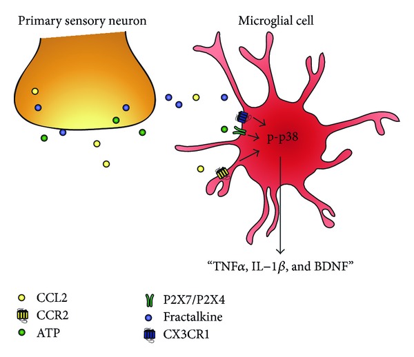
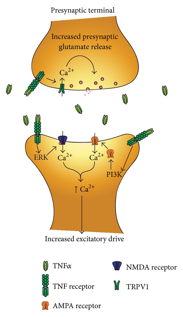
Microglia are regarded as macrophages in the central nervous system (CNS) and play an important role in neuroinflammation in the CNS. Microglial activation has been strongly implicated in neurodegeneration in the brain. Increasing evidence also suggests an important role of spinal cord microglia in the genesis of persistent pain, by releasing the proinflammatory cytokines tumor necrosis factor-alpha (TNFα), Interleukine-1beta (IL-1β), and brain derived neurotrophic factor (BDNF). In this review, we discuss the recent findings illustrating the importance of microglial mediators in regulating synaptic plasticity of the excitatory and inhibitory pain circuits in the spinal cord, leading to enhanced pain states. Insights into microglial-neuronal interactions in the spinal cord dorsal horn will not only further our understanding of neural plasticity but may also lead to novel therapeutics for chronic pain management.


Figures

Figure 1
Figure 1
Nerve injury induces marked microglial reaction in the ipsilateral lumbar spinal cord 7 days after chronic constriction injury (CCI). Microglia as demonstrated by CX3CR1 expression in CX3CR1-GFP mice. The contralateral side (left) shows the typical resting microglial morphology, and the ipsilateral side (right) illustrates the enlarged and amoeboid morphological features of activated microglia cell bodies. Also note that on the ipsilateral side the number and density of microglia in the medial superficial dorsal horn and lateral ventral horn, where injured primary afferent axons terminate, are significantly increased. Scale, 100 μm.
Figure 2
Figure 2
Schematic of primary afferent releasing factors that are responsible for microglial activation. Microglial cells are activated by factors released from hyperexcitable primary afferents such as CCL2 (MCP-1), ATP, and CX3CL1 (fractalkine). Respective activation of CCR2, P2X4/P2X7, and CX3CR1 receptors on microglia causes phosphorylation of p38 in microglia, leading to increased synthesis and release of TNFα, IL-1β, and BDNF.
Figure 3
Figure 3
Schematic of TNFα induced potentiation of spinal cord synaptic transmission. Microglial release of TNFα increases excitatory neurotransmission in the dorsal horn via both presynaptic and postsynaptic mechanisms. At presynaptic sites, TNFα increases glutamate release via TRPV1 activation and there will be a subsequent increase in intracellular Ca2+. At postsynaptic sites, TNFα increases the activity of AMPA and NMDA receptors via activation of PI3 K and ERK on glutamatergic neurons to increase excitatory drive.

References

    1. Ransohoff RM, Perry VH. Microglial physiology: unique stimuli, specialized responses. Annual Review of Immunology. 2009;27:119–145. - PubMed
    1. Lawson LJ, Perry VH, Dri P, Gordon S. Heterogeneity in the distribution and morphology of microglia in the normal adult mouse brain. Neuroscience. 1990;39(1):151–170. - PubMed
    1. Perry VH, Gordon S. Macrophages and the nervous system. International Review of Cytology. 1991;125:203–244. - PubMed
    1. Zhang F, Vadakkan KI, Kim SS, Wu L-J, Shang Y, Zhuo M. Selective activation of microglia in spinal cord but not higher cortical regions following nerve injury in adult mouse. Molecular Pain. 2008;4, article 15 - PMC - PubMed
    1. Ayoub AE, Salm AK. Increased morphological diversity of microglia in the activated hypothalamic supraoptic nucleus. Journal of Neuroscience. 2003;23(21):7759–7766. - PMC - PubMed

Publication types

LinkOut - more resources