Review
. 2013 Jan 19;1(1):140-4.
doi: 10.1016/j.redox.2013.01.006.

Lipofuscin: formation, effects and role of macroautophagy

Affiliations
Review

Lipofuscin: formation, effects and role of macroautophagy

Annika Höhn et al. Redox Biol. .

Abstract

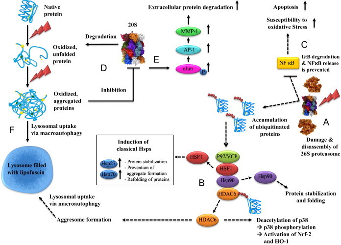
Oxidative stress plays a crucial role in the development of the aging process and age dependent diseases. Both are closely connected to disturbances of proteostasis by protein oxidation and an impairment of the proteasomal system. The final consequence is the accumulation of highly cross-linked undegradable aggregates such as lipofuscin. These aggregates of damaged proteins are detrimental to normal cell functions. Here we provide an overview about effect of these aggregates on the proteasomal system, followed by transcription factor activation and loss of cell viability. Furthermore, findings on the mechanism of radical genesis, proteasomal inhibition and the required components of lipofuscin formation were resumed.

Keywords: Aging; Autophagy; Lipofuscin; Oxidative stress; Proteasome.


Figures

Fig. 1
Fig. 1
Consequences of oxidative stress on proteins, the proteasomal system and transcription factors. (A) The 26S proteasome is highly susceptible to inactivation during oxidative stress (figured as flash symbol) leading to an excessive accumulation of polyubiquitinated proteins. (B) Histone deacetylase 6 (HDAC6) has the ability to sense and bind ubiquitinated proteins via the BUZ (bound to ubiquitin zinc finger) domain which induces the dissociation of a complex formed out of Hsp90, HSF-1, p97/VCP and HDAC6 . Following the liberation of HDAC6 and p97/VCP , the latter uses either its ATP dependent segregase activity to dissolve the bond between Hsp90 and HSF-1 directly or p97/VCP stimulates the Hsp90 ATPase activity, which results in the release of HSF-1 and the consequence up-regulation of several heat shock proteins, such as Hsp70 and Hsp 27 . Beyond up-regulation of classical Hsps upon proteasome inhibition, HDAC6 is also involved in an induction of HO-1 after proteasome inhibition . Initiation of this pathway is also the detection of ubiquitinated proteins and the release of HDAC6 and mediated by a p38/MAPK-dependent activation of Nrf-2, which is the most important transcriptional activator of HO-1 gene translation [171]. Further HDAC6 has the ability to favor the accumulation of polyubiquitinated proteins in cellular aggresomes by interaction with ubiquitin and dynein motors. Aggresomes are inclusion bodies next to the nucleus at the proximity end of the microtubule organizing center, finally eliminated by an autophagy-mediated mechanism. (C) Furthermore, a pivotal regulatory protein function is disrupted by 26S proteasomal inhibition, the transcription factor NFϰB. This factor binds to multiple DNA sequences, initiating the transcription of gene products including various cytokines, angiogenesis factors, cell adhesion molecules, enzymes and antiapoptotic factors . NFϰB is located in the cytoplasm in an inactive form, bound to an inhibitor molecule IϰB. Stimulation of cells through a variety of mechanisms triggers a cascade of signaling events resulting in the degradation of IϰB by the proteasome. This degradation releases active NFϰB, which then translocates into the nucleus and binds to specific DNA sequences on its target genes . Proteasomal inhibition blocks NFϰB activation and leads among others to increased susceptibility to oxidative stress and apoptosis. (D) The activity of the 20S proteasome, which is mostly responsible for the degradation of oxidized proteins and less susceptible to direct oxidative stress, can be nevertheless diminished by aggregated oxidized proteins. These protein aggregates are formed under stress conditions as complexes of unfolded proteins which do not normally interact with each other. It might require several steps depending on the nature of the initial conditions, leading to unfolding and aggregate formation. Due to the complex process of intermolecular interactions, such as during physiological aging, the process of aggregation is slow . The aggregate is independent from the original structure of the protein and introduces a new toxic element into cellular metabolism, partly by inhibiting 20S proteasome. (E) Another side effect of proteasome inhibition is leading to a higher amount of phosphorylated c-Jun leading to an activation of AP-1, known to control the expression of MMP-1 and numerous other genes. Subsequently increased extracellular protein degradation can be assumed, as observed in skin aging. (F) When proteolytic capacity declines below a critical threshold of activity required to cope with oxidative stress, the final consequence is the accumulation of aggregated proteins which may, instead, cross-link with one another or form extensive hydrophobic bonds. This material might undergo further reactions and finally form the age pigment lipofuscin which has toxic properties and accumulates in the lysosomal system..
Fig. 2
Fig. 2
Properties of lipofuscin. Studies have suggested that lipofuscin is not an inert waste product, but rather an active component influencing the cellular metabolism, which is especially relevant in senescent cells. It was proposed that lipofuscin is cytotoxic because of its ability to incorporate redox-active transition metals, resulting in a redox-active surface, able to catalyze the Fenton reaction: in the presence of Fe2+, H2O2 is decomposed forming hydroxyl radicals (OH) . The resulting Fe3+ can be reduced by superoxide and the vicious cycle starts again. This ability to incorporate transition metals (iron) and form oxidants was tested by using artificial lipofuscin-like oxidized, cross-linked protein aggregates (“artificial lipofuscin”). Non-iron-loaded artificial lipofuscin was incubated with Fe2+ and was able to incorporate a maximum amount of 7 mass%, matching the characteristics of native lipofuscin very well . In vitro this material is able to generate the formation of free radicals and initiates apoptotic cell death, resulting in a significant loss of cellular viability (A). Another major and already demonstrated characteristic of protein aggregates/lipofuscin is the ability to inhibit the degradation of oxidized proteins by competitively binding to the proteasome [40–42]. The proposed mechanism for proteasomal inhibition is binding to exposed hydrophobic amino acid structures on the surface of the highly oxidized and covalently cross-linked lipofuscin . These structures seem to be main sites of proteasomal substrate recognition. After proteasomal binding to those structures the protease is unable to degrade the exposed structures completely because of sterical and/or mechanical inhibition by cross-links. So the proteasome is bound to the surface of lipofuscin in ineffective attempts of degradation detracting proteolytic capacity from other substrates and resulting in a measurable proteasomal inhibition. Furthermore, celluar viability is affected and significantly reduced (B). Degradation of these exposed structures by using the effective and nonspecific protease K demonstrated that such unfolded peptide binding sites are largely responsible for the inhibition of the proteasome. This degradation of these binding sites significantly reduces the proteasomal binding and the inhibition of the proteasome could be partially prevented. Consequently less proteasomal capacity is detracted in futile attempts of degradation and cellular viability was unaffected or increased referred to the undegraded material (C) .
Fig. 3
Fig. 3
The fate of aggregated proteins during functional macroautophagy. Macroautophagy is a process, which is responsible for the uptake of larger aggregates. Aggregates which are no longer degradable by the proteasome, but rather inhibit its proteolytic function are enclosed by a phagophore and transported towards the lysosomal system. Macroautophagy comprise the formation of an autophagosome, a double-membrane vesicle that engulfs substrates trough the expansion of an isolation membrane, called phagophore. This isolation membrane is build, expanded and closed to an autophagosome and delivered to the endo/lysosomal system. Afterwards both structures fuse and the final structure is called autophagolysosome. The elongation of the phagophore requires two processes similar to ubiquitination: Atg12-Atg5-conjugation and LC3 (microtubule-associated-protein-light-chain-3)-modification (illustration of Atg12-Atg5-system and LC3 conjugation system according to Levine and Deretic [43]). In the first conjugation system (Atg12-Atg5-system) Atg12 is conjugated with Atg5, in a process assisted by an E1-ubiquitin-like activating enzyme: Atg7 and the E2-ubiquitin-like conjugation enzyme: Atg10. Afterwards the Atg12-Atg5 conjugate forms a protein complex with Atg16 on the outside of the nascent autophagosome . This complex is functionally necessary for the end product of the secondary conjugation system, lipidated LC3. First the cysteine protease Atg4 eliminates the C-terminal tail of LC3I to expose a glycine residue. A G116 is activated by Atg7, and LC3I is conjugated via a thioester bond with Atg7. Afterwards LC3I is transferred on the E2-ubiquitin-like conjugation enzyme Atg3, and again connected via a thioester bond. Finally LC3I is bound to the amino group of phosphatidylethanolamine (PE). This complex of LC3 with PE is called LC3II and localized via its lipid part to the autophagosomal membrane enabling membrane-elongation. Finally LC3II is deconjugated from the phospholipid-anchor via Atg4 and the luminal associated LC3II is degraded. This cycle of conjugation and deconjugation is important for the normal progression of autophagy. Especially under conditions of oxidative stress a huge amount of aggregated proteins accumulate. These aggregates are taken up by macroautophagy and react further with other cellular components, forming a fluorescent material referred to as lipofuscin, which accumulates in the lysosomal system over time.
Fig. 4
Fig. 4
The fate of aggregated proteins during impaired or non-sufficient macroautophagy. Macroautophagy is maximally activated under stress conditions such as starvation, oxidative stress and conditions leading to enhanced protein misfolding [46–50]. This essential role of macroautophagy is underpinned by the fact that Atg5 or Atg7 null mice die a few hours after birth . Nevertheless it is possible to investigate the role of macroautophagy in knockout models such as fibroblasts from mouse embryos deficient in Atg5 (Atg5−/− MEFs) under stress conditions. This ATG5−/− MEFs naturally show no Atg5 expression and thus no formation of the Atg12-Atg5-complex. As the Atg12-Atg5-conjugation system and LC3 modification (illustration according to Levine and Deretic [43]) are both mandatory for the formation of a phagophore, the uptake of aggregated proteins (and other substrates) is prevented in this knockdown system. The consequence is an increasing amount of cytosolic protein aggregates and finally a dramatically enhanced cytosolic lipofuscin accumulation. These elevated levels of extralysosomal lipofuscin are combined with a decline in cellular viability and an increased amount of ROS production.

References

    1. Morimoto R.I. Proteotoxic stress and inducible chaperone networks in neurodegenerative disease and aging. Genes Development. 2008;22:1427–1438. - PMC - PubMed
    1. Pickering A.M., Koop A.L., Teoh C.Y., Ermak G., Grune T., Davies K.J. The immunoproteasome, the 20S proteasome and the PA28alphabeta proteasome regulator are oxidative-stress-adaptive proteolytic complexes. Biochemical Journal. 2010;432:585–594. - PMC - PubMed
    1. Ullrich O., Reinheckel T., Sitte N., Hass R., Grune T., Davies K.J. Poly-ADP ribose polymerase activates nuclear proteasome to degrade oxidatively damaged histones. Proceedings of the National Academy of Sciences USA. 1999;96:6223–6228. - PMC - PubMed
    1. Grune T., Reinheckel T., Davies K.J. Degradation of oxidized proteins in K562 human hematopoietic cells by proteasome. Journal of Biological Chemistry. 1996;271:15504–15509. - PubMed
    1. Pacifici R.E., Kono Y., Davies K.J. Hydrophobicity as the signal for selective degradation of hydroxyl radical-modified hemoglobin by the multicatalytic proteinase complex, proteasome. Journal of Biological Chemistry. 1993;268:15405–15411. - PubMed