. 2013 Feb 22;1(1):265-75.
doi: 10.1016/j.redox.2013.02.001. eCollection 2013.

Acrolein-induced activation of mitogen-activated protein kinase signaling is mediated by alkylation of thioredoxin reductase and thioredoxin 1

Affiliations

Acrolein-induced activation of mitogen-activated protein kinase signaling is mediated by alkylation of thioredoxin reductase and thioredoxin 1

Matthew J Randall et al. Redox Biol. .

Abstract

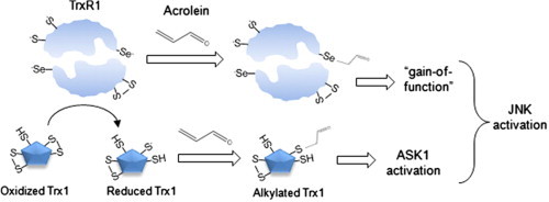
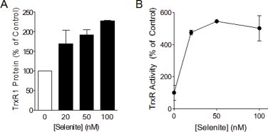
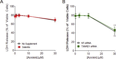
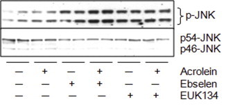
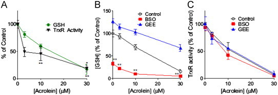
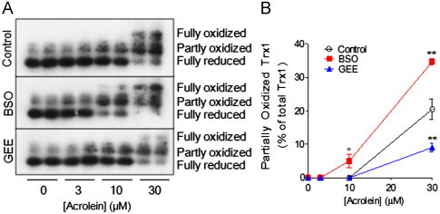
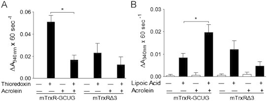
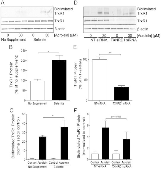
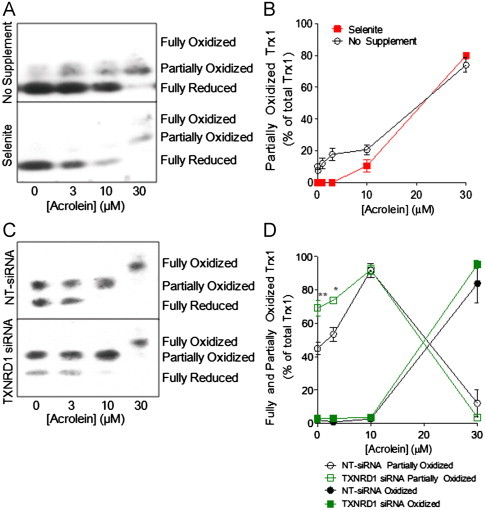
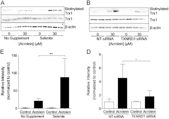
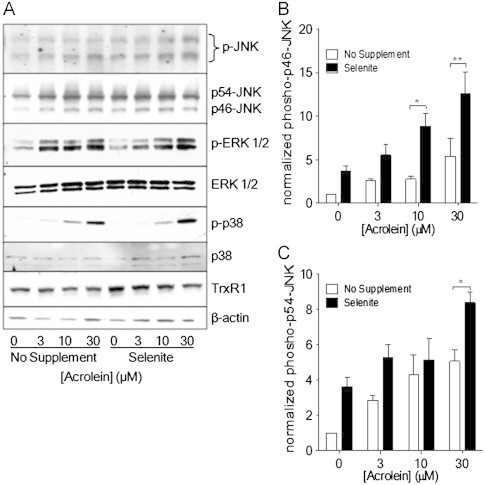
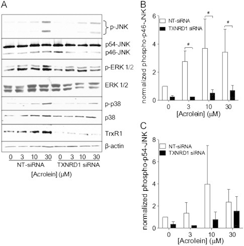
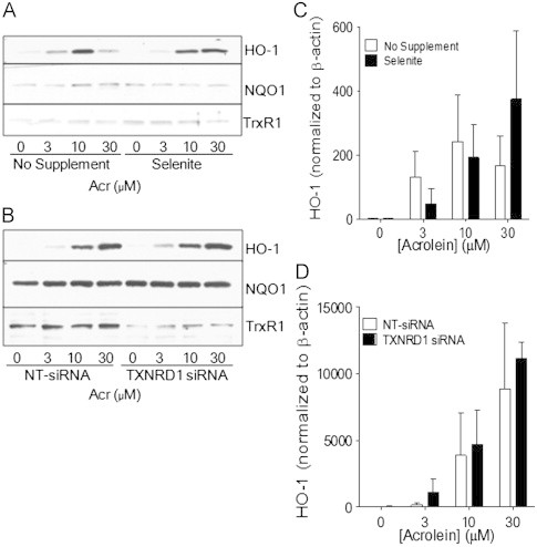
Cigarette smoking remains a major health concern worldwide, and many of the adverse effects of cigarette smoke (CS) can be attributed to its abundant electrophilic aldehydes, such as acrolein (2-propenal). Previous studies indicate that acrolein readily reacts with thioredoxin reductase 1 (TrxR1), a critical enzyme involved in regulation of thioredoxin (Trx)-mediated redox signaling, by alkylation at its selenocysteine (Sec) residue. Because alkylation of Sec within TrxR1 has significant implications for its enzymatic function, we explored the potential importance of TrxR1 alkylation in acrolein-induced activation or injury of bronchial epithelial cells. Exposure of human bronchial epithelial HBE1 cells to acrolein (1-30 μM) resulted in dose-dependent loss of TrxR thioredoxin reductase activity, which coincided with its alkylation, as determined by biotin hydrazide labeling, and was independent of initial GSH status. To test the involvement of TrxR1 in acrolein responses in HBE1 cells, we suppressed TrxR1 using siRNA silencing or augmented TrxR1 by cell supplementation with sodium selenite. Acrolein exposure of HBE1 cells induced dose-dependent activation of the MAP kinases, extracellular regulated kinase (ERK), c-Jun N-terminal kinase (JNK), and p38, and activation of JNK was markedly enhanced after selenite-mediated induction of TrxR1, and was associated with increased alkylation of TrxR1. Conversely, siRNA silencing of TrxR1 significantly suppressed the ability of acrolein to activate JNK, and also appeared to attenuate acrolein-dependent activation of ERK and p38. Alteration of initial TrxR1 levels by siRNA or selenite supplementation also affected initial Trx1 redox status and acrolein-mediated alkylation of Trx1, but did not significantly affect acrolein-mediated activation of HO-1 or cytotoxicity. Collectively, our findings indicate that alkylation of TrxR1 and/or Trx1 may contribute directly to acrolein-mediated activation of MAP kinases such as JNK, and may therefore be important in acrolein-induced alterations in airway epithelial function, as a contributing mechanism in tobacco-related respiratory disease.

Keywords: Cigarette smoke; Electrophile; Epithelium; GSH; Michael addition; c-Jun N-terminal kinase.


Figures

None
Graphical abstract
Fig. S1
Fig. S1
Selenite supplementation alters TrxR1 protein levels and enzyme activity. HBE1 cells were supplemented with selenite (20–100 nM) for a duration of 5 days after which cells were lysed for analysis of TrxR1 protein levels by Western blotting (A), and TrxR activity using the insulin assay as described in the Methods section (B). Results are expressed as mean±SEM.
Fig. S2
Fig. S2
TrxR1 status does not affect acrolein-induced toxicity. Selenite-supplemented (A) or TXNRD1 siRNA transfected (B) HBE1 cells were treated with acrolein, and cell viability was assessed by analysis of LDH release in the culture media after 24 h. Results are expressed as mean±SEM, n=3. **: p<0.01 compared to control.
Fig. S3
Fig. S3
Effect of ROS scavengers on acrolein-mediated JNK phosphorylation. HBE1 cells were pretreated for 30 min with either ebselen (10 μM) or EUK134 (50 μM) followed by a 30 min exposure of acrolein (30 μM), after which cells were lysed and p-JNK and JNK were analyzed by Western blot. Representative blots of two experiments are shown.
Fig. 1
Fig. 1
Acrolein-induced redox alterations in HBE1 cells. HBE1 cells were treated with acrolein at indicated concentrations for 30 min after which TrxR activity and GSH levels were measured as described in the Methods section (A). HBE1 cells were pretreated with BSO (24 h) or GEE (4 h) prior to acrolein treatment of indicated for 30 min, after which GSH levels (B) and TrxR1 activity (C) were measured. Results are expressed as mean±SEM, n=4. *: p<0.05, **: p<0.01 compared to control.
Fig. 2
Fig. 2
Acrolein-dependent Trx oxidation depends on GSH status. HBE1 cells were pretreated with BSO (24 h) or GEE (4 h) prior to acrolein treatment for 30 min, and Trx1 redox status was determined by S-carboxymethylation and non-reducing native PAGE. Representative redox Western blots are shown in (A) and relative band densities of partially oxidized Trx are summarized in (B). Results are expressed as mean±SEM, n=4. *: p<0.05, **: p<0.01 compared to control.
Fig. 3
Fig. 3
Effects of acrolein on enzymatic activity of mTrxR. Semisynthetic mTrxR-GCUG and mTrxRΔ3 (200 nM) were incubated with acrolein for 30 min, and thioredoxin reductase activity was assessed using the insulin assay (A), and NADPH oxidase activity was determined in the absence (white bars) or presence (black bars) of α-lipoic acid (B). Results expressed as the mean change in absorbance x 60 s−1±SEM, n=5. *: p<0.05 compared to control enzyme.
Fig. 4
Fig. 4
Alterations in TrxR by selenite supplementation or siRNA silencing. HBE1 cells were supplemented with 50 nM selenite (A, B and C) or transfected with TXNRD1 or NT siRNA (D, E and F), and treated with 30 μM acrolein for 30 min for Western blot analysis of total TrxR1 protein levels or β-actin in whole cell lysates (B and E), or analysis of acrolein-modified TrxR1 which was determined after labeling with biotin hydrazide and analysis of avidin-purified proteins by Western blot for TrxR1 (C and F). Densitometry of Western blots was analyzed and results are expressed as mean±SEM. *: p<0.05, **: p<0.01 compared to non-supplemented or NT-siRNA controls.
Fig. 5
Fig. 5
Effects of TrxR1 alterations on acrolein-induced Trx1 oxidation/alkylation. HBE1 cells were supplemented with selenite or transfected with TXNRD1 siRNA and treated with acrolein for 30 min, and the oxidation/alkylation status of Trx1 was determined by redox Western blot (A and C). Graphs show relative band densities of partially and/or fully oxidized Trx1 (B and D). Results are expressed as mean±SEM, n=4. *: p<0.05, **: p<0.01 non-supplemented or NT-siRNA controls.
Fig. 6
Fig. 6
Alterations in Trx1 by selenite supplementation or siRNA silencing. HBE1 cells were supplemented with 50 nM selenite (A) or transfected with TXNRD1 or NT siRNA (B), and treated with 30 μM acrolein for 30 min for Western blot analysis of total Trx1 protein levels or β-actin in whole cell lysates, or analysis of acrolein-modified TrxR1 which was determined after labeling with biotin hydrazide and analysis of avidin-purified proteins by Western blot for Trx1. Graphs show relative band densities of Biotinylated Trx1 (C, D). Results are expressed as mean±SEM. *: p<0.05, **: p<0.01 compared to non-supplemented or NT-siRNA controls.
Fig. 7
Fig. 7
Effect of selenite supplementation on acrolein-induced MAPK activation. Non-supplemented or selenite-supplemented HBE1 cells were treated with indicated concentrations of acrolein for 30 min, and cell lysates were analyzed for p-JNK, p-ERK1/2, and p-p38 by Western blot (A). Band densities of phosphorylated p46-JNK (B) and p56-JNK (C) were quantified relative to total JNK protein. Results are expressed as mean±SEM, n=3. *: p<0.05, **: p<0.01 compared to non-supplemented controls.
Fig. 8
Fig. 8
Effect of TrxR1 siRNA silencing on acrolein-induced MAPK activation. HBE1 cells were transfected with Non-targeting (NT-siRNA) or TXNRD1 siRNA and treated with 30 μM acrolein for 30 min, and cell lysates were analyzed for p-JNK, p-ERK, and p-p38 by Western Blot (A). Band densities of phosphorylated p46-JNK (B) and p54-JNK (C) were quantified relative to total JNK protein. Results expressed as mean±SEM, n=3. *: p<0.05 compared to NT-siRNA controls.
Fig. 9
Fig. 9
Effect of TrxR1 status on acrolein-induced phase II responses. Selenite-supplemented (A) or TXNRD1 siRNA transfected (B) HBE1 cells were treated with acrolein and expression of HO-1, NQO-1, and TrxR1 were analyzed after 8 h by Western blot. Quantified HO-1 band densities relative to β-actin are presented as mean±SEM (n=4) (C, D).

References

    1. Office of the Surgeon General. The Health Consequences of Smoking: A Report of the Surgeon General; 2004. - PubMed
    1. Saetta M. Airway inflammation in chronic obstructive pulmonary disease. American Journal of Respiratory and Critical Care Medicine. 1999;160:S17–20. - PubMed
    1. Thomson N.C., Chaudhuri R. Asthma in smokers: challenges and opportunities. Current Opinion in Pulmonary Medicine. 2009;15:39–45. - PubMed
    1. Heijink I.H., Brandenburg S.M., Postma D.S., van Oosterhout A.J. Cigarette smoke impairs airway epithelial barrier function and cell–cell contact recovery. European Respiratory Journal. 2012;39:419–428. - PubMed
    1. Xiao C., Puddicombe S.M., Field S., Haywood J., Broughton-Head V., Puxeddu I., Haitchi H.M., Vernon-Wilson E., Sammut D., Bedke N., Cremin C., Sones J., Djukanovic R., Howarth P.H., Collins J.E., Holgate S.T., Monk P., Davies D.E. Defective epithelial barrier function in asthma. Journal of Allergy and Clinical Immunology. 2011;128:549–556. e541-512. - PubMed

Publication types