. 2014 Feb 13;57(3):539-66.
doi: 10.1021/jm400674a. Epub 2013 Sep 25.

Inhibiting the HIV integration process: past, present, and the future

Affiliations

Inhibiting the HIV integration process: past, present, and the future

Roberto Di Santo. J Med Chem. .

Erratum in

  • J Med Chem. 2014 Jul 24;57(14):6273

Abstract

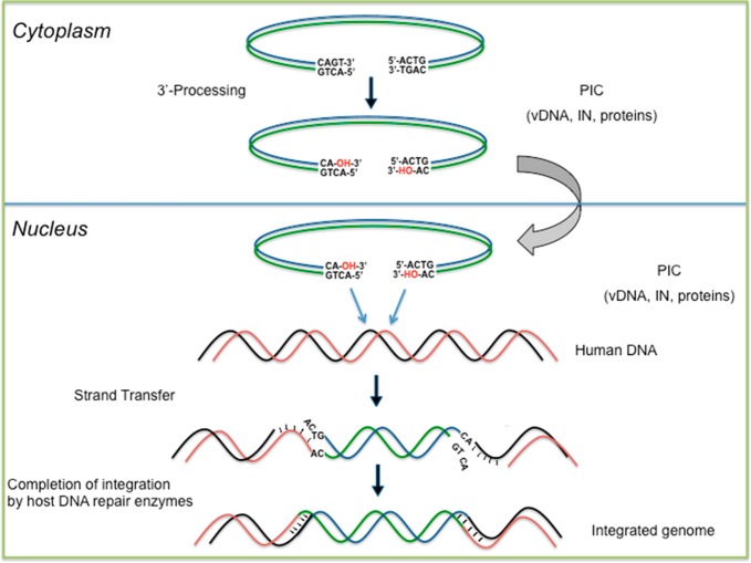
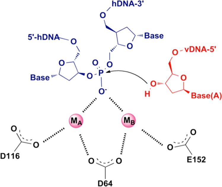
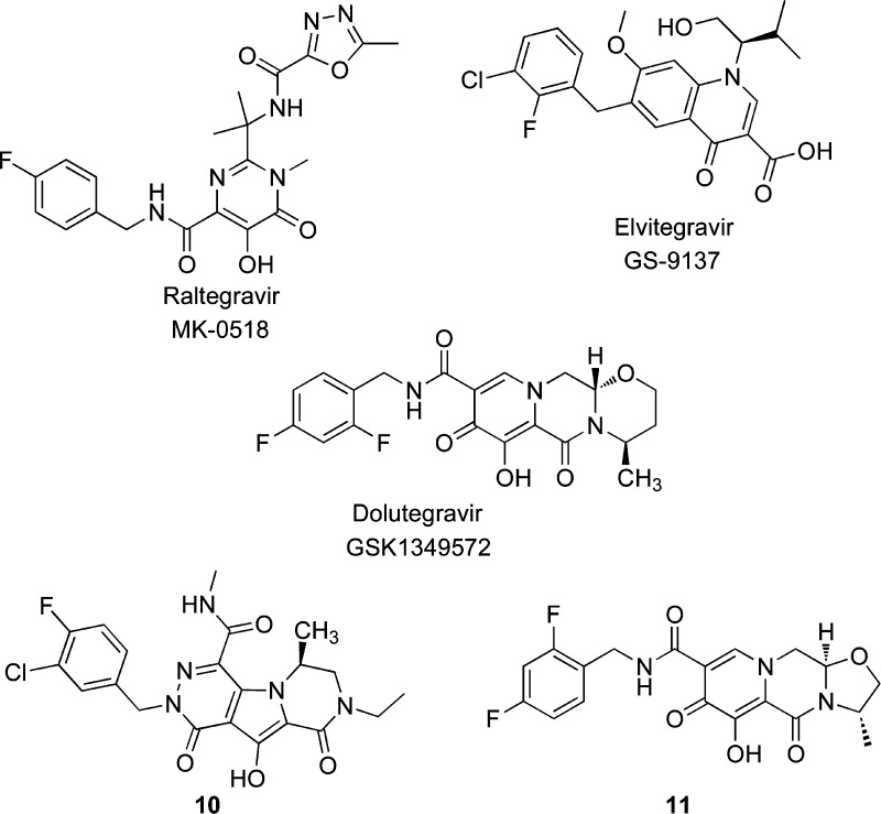
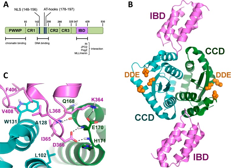
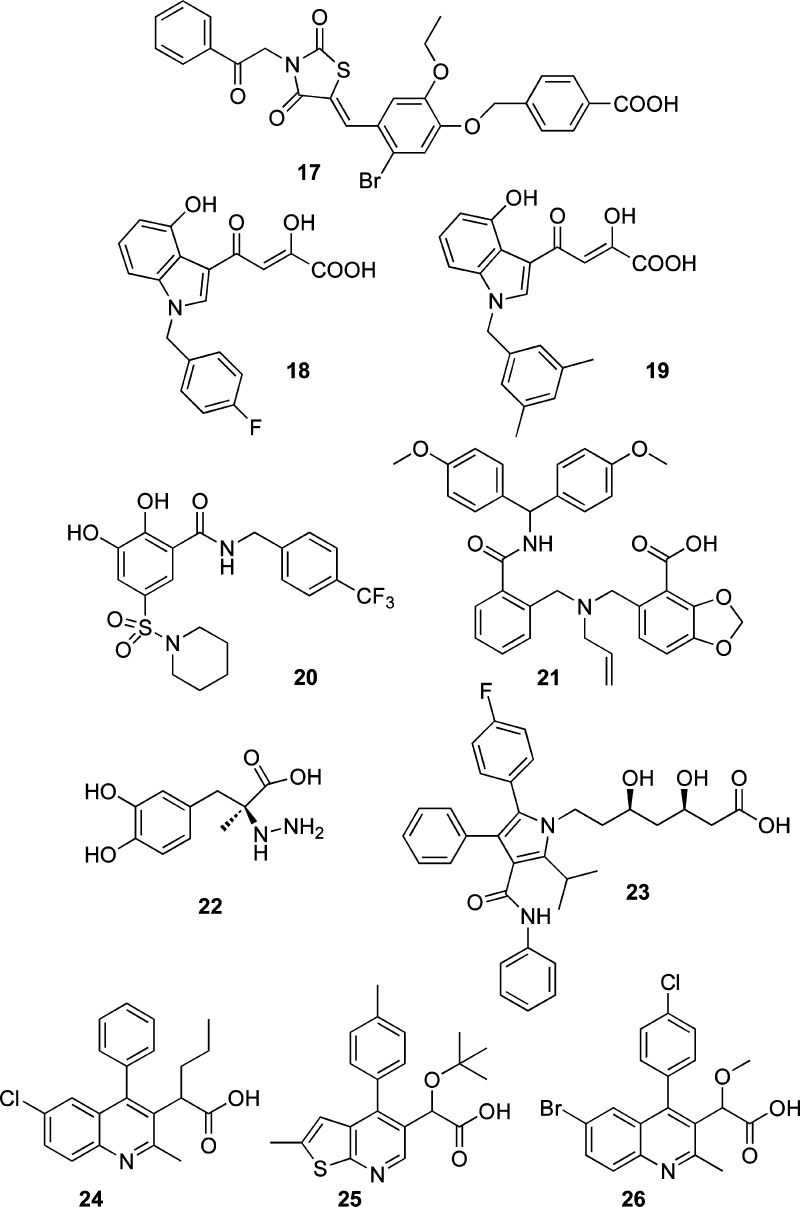
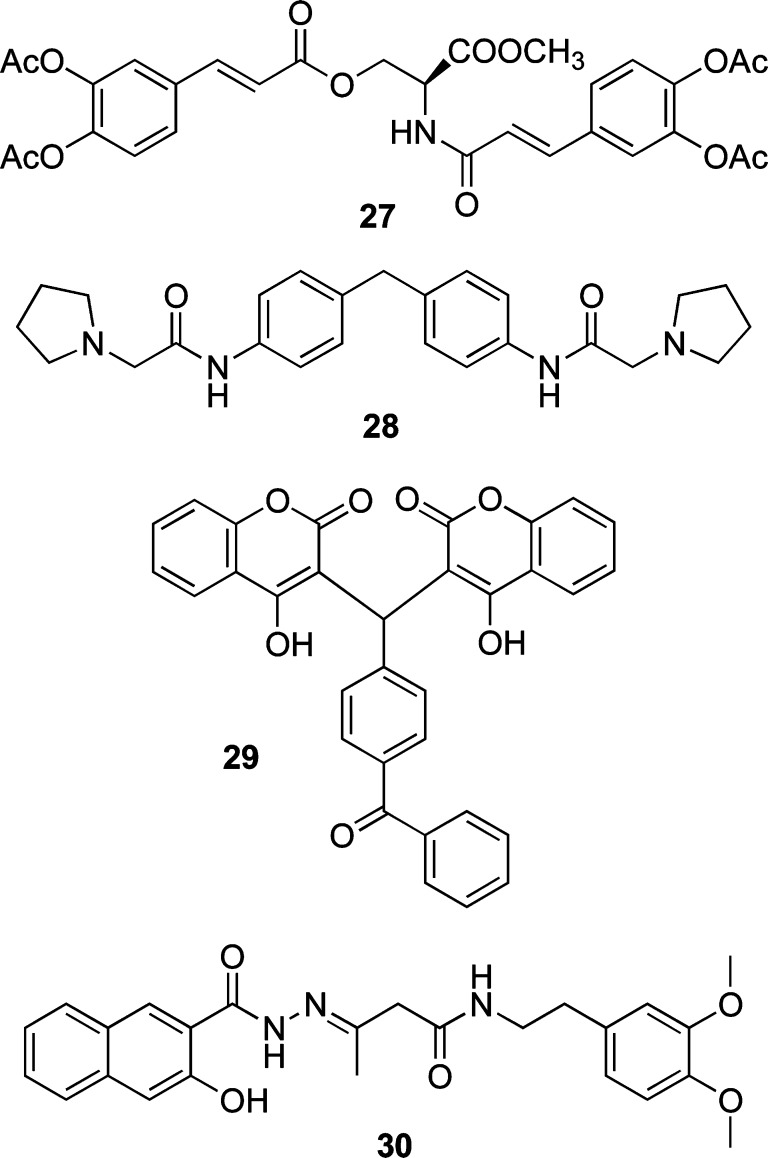
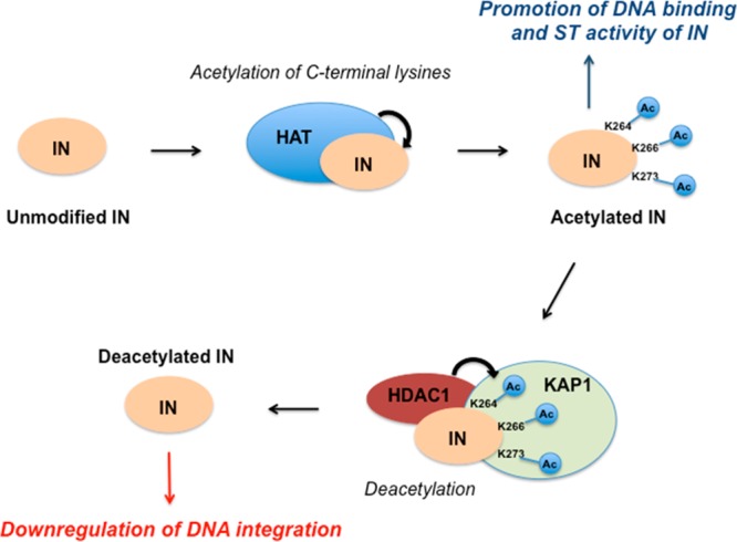
HIV integrase (IN) catalyzes the insertion into the genome of the infected human cell of viral DNA produced by the retrotranscription process. The discovery of raltegravir validated the existence of the IN, which is a new target in the field of anti-HIV drug research. The mechanism of catalysis of IN is depicted, and the characteristics of the inhibitors of the catalytic site of this viral enzyme are reported. The role played by the resistance is elucidated, as well as the possibility of bypassing this problem. New approaches to block the integration process are depicted as future perspectives, such as development of allosteric IN inhibitors, dual inhibitors targeting both IN and other enzymes, inhibitors of enzymes that activate IN, activators of IN activity, as well as a gene therapy approach.


Figures

Figure 1
Figure 1
Structure of HIV-1 IN. (A) IN domains, where the catalytic triad is shown in pink. (B–D) Structures of single IN domains: (B) NTD (PDB code 1wje); (C) CCD (PDB code 1bis); (D) CTD (PDB code 1ihv). (E–F) IN two-domain structures: (E) NTD + CCD (PDB code 1k6y); (F) CTD + CCD (PDB code 1ex4). Each structure consists of two IN monomers, shown in yellow and blue. The zinc ions in the NTD are shown in green, and the catalytic triad (D64, D116, and E152) in the CCD is shown in pink.
Figure 2
Figure 2
Architecture of the PFV intasome. (A) Views along (left) and perpendicular (right) to the crystallographic 2-fold axis. The subunits of the IN tetramer, which are in blue and green, are engaged with viral DNA. The external IN chains are in yellow. The DNA strands are orange and magenta, and the last one is the most reactive. D128, D185, E221, or the catalytic triad is in red. Gray spheres are Zn atoms. (B) Focus on IN chains with domains and linkers indicated.
Figure 3
Figure 3
Outline of the in vivo integration process.
Figure 4
Figure 4
ST step. The attack of the 3′ ends of vDNA on the phosphodiester bonds of host DNA is coordinated by metal ions.
Chart 1
Chart 1. DKAs and Bioisosteres as IN Selective ST Inhibitors
Figure 5
Figure 5
Design of early RAL-like inhibitors: from HCV polymerase to HIV IN inhibitors.
Chart 2
Chart 2. First and Second Generation of the INSTIs Approved for Clinical Trials
Chart 3
Chart 3. Dual Inhibitors of IN Enzyme and RNase H Function of RT
Figure 6
Figure 6
Representative list of IN- and PIC-associated viral (blue) and cellular (red) proteins in retroviral replication.
Figure 7
Figure 7
(A) LEDGF/p75 domains: N-terminal PWWP motif and the charged regions (CR1–3) critical for chromatin recognition and the central DNA binding domain (blue) and the C-terminal IBD (magenta) essential for binding to IN and cellular proteins. (B) Cocrystallized structure of LEDGF/p75–IBD (magenta) and the CCD dimer of integrase (green and blue). The catalytic triad is represented in orange (PDB code 2B4J). (C) Cartoon focused on CCD–IBD binding (PDB code 2B4J). IN CCDs are shown in green and blue, whereas the LEDGF/p75 IBD is in magenta. Residues of IN (dark green) and IBD (magenta) critical for the interaction are highlighted.
Figure 8
Figure 8
Schematic representation of the LEDGF support of the HIV-1 integration process. On the right, the different mechanisms of inhibition by LEDGINs (up) and INSTIs (down) are described. Inhibitors are represented in red.
Chart 4
Chart 4. Small Molecule Inhibitors of the LEDGF/p75–IN Interaction
Chart 5
Chart 5. Small Molecule Inhibitors of IN Multimerization
Figure 9
Figure 9
Regulation of HIV-1 integration by the acetylation and deacetylation of IN.

Similar articles

Cited by

References

    1. Gottlieb M. S.; Schroff R.; Schanker H. M; Weisman J. D.; Fan P. T; Wolf R. A.; Saxon A. Pneumocystis carinii pneumonia and mucosal candidiasis in previously healthy homosexual men: evidence of a new acquired cellular immunodeficiency. N. Engl. J. Med. 1981, 305, 1425–1431. - PubMed
    1. Masur H.; Michelis M. A.; Greene J. B.; Onorato I.; Stouwe R. A.; Holzman R. S.; Wormser G.; Brettman L.; Lange M.; Murray H. W.; Cunningham-Rundles S. An outbreak of community-acquired Pneumocystis carinii pneumonia: initial manifestation of cellular immune dysfunction. N. Engl. J. Med. 1981, 305, 1431–1438. - PubMed
    1. Siegal F. P.; Lopez C.; Hammer G. S.; Brown A. E.; Kornfeld S. J.; Gold J.; Hassett J.; Hirschman S. Z.; Cunningham-Rundles C.; Adelsberg B. R.; Parham D. M.; Siegal M.; Cunningham-Rundles S.; Armstrong D. Severe acquired immunodeficiency in male homosexuals, manifested by chronic perianal ulcerative herpes simplex lesions. N. Engl. J. Med. 1981, 305, 1439–1444. - PubMed
    1. Barton K. M.; Burch B. D.; Soriano-Sarabia N.; Margolis D. M. Prospects for treatment of latent HIV. Clin. Pharmacol. Ther. 2013, 93, 46–56. - PMC - PubMed
    1. Cohen J. Therapies. Confronting the limits of success. Science 2002, 296, 2320–2324. - PubMed

Publication types

MeSH terms