. 2013 Sep 11;12(1):105.
doi: 10.1186/1476-4598-12-105.

miR-204 mediated loss of Myeloid cell leukemia-1 results in pancreatic cancer cell death

Affiliations

miR-204 mediated loss of Myeloid cell leukemia-1 results in pancreatic cancer cell death

Zhiyu Chen et al. Mol Cancer. .

Abstract

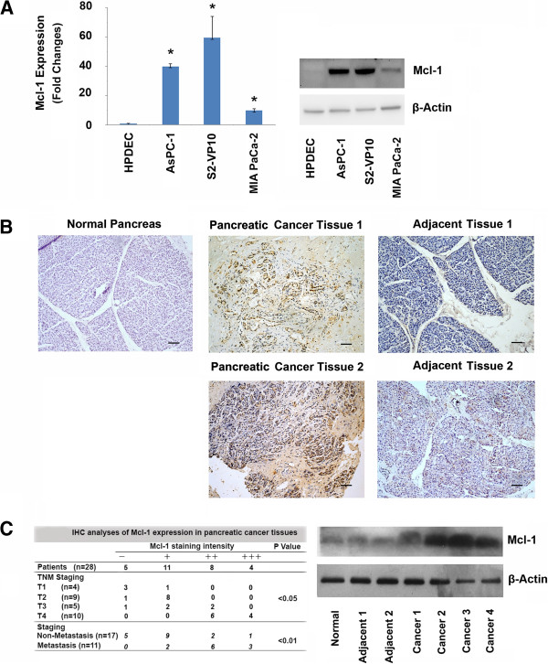
Background: Pancreatic cancer is one of the most lethal human malignancies, with an all-stage 5-year survival of <5%, mainly due to lack of effective available therapies. Cancer cell survival is dependent upon up-regulation of the pro-survival response, mediated by anti-apoptotic proteins such as Mcl-1.

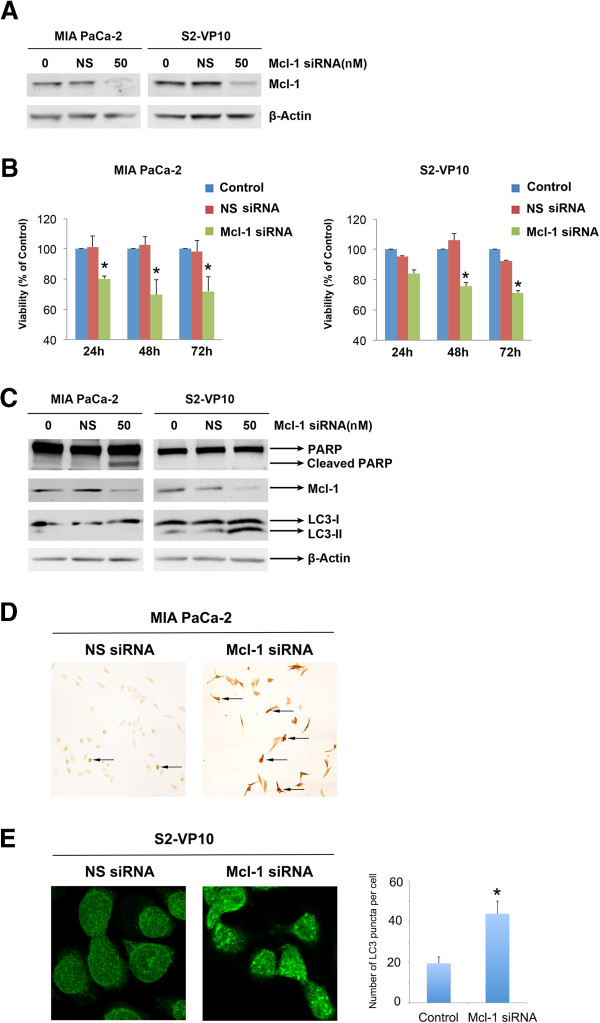
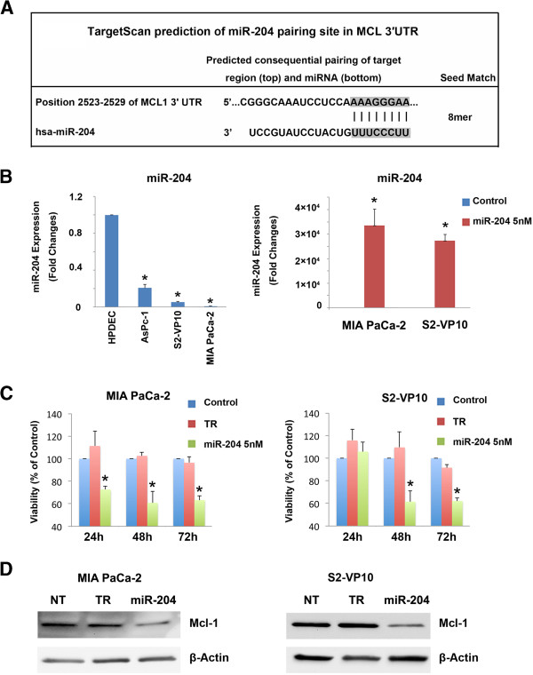
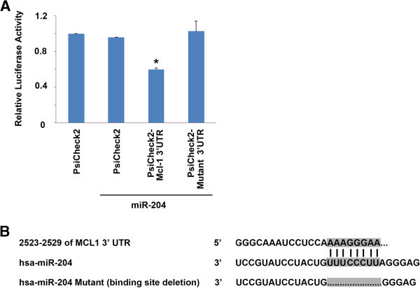
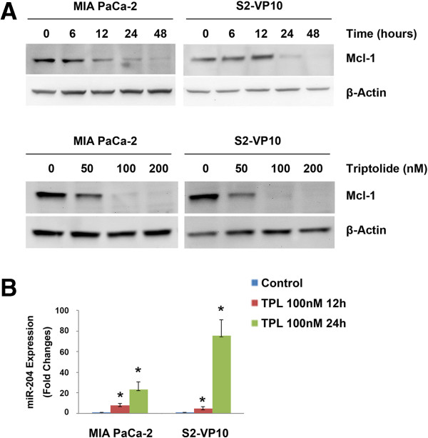
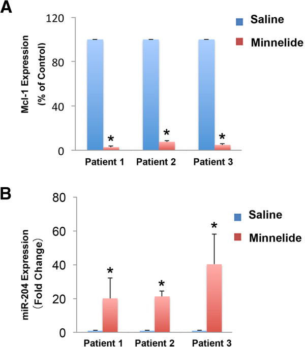
Results: Here we show that over-expression of Mcl-1 in pancreatic patient tumor samples is linked to advancement of the disease. We have previously shown that triptolide, a diterpene triepoxide, is effective both in vitro and in vivo, in killing pancreatic cancer cells. Decrease of Mcl-1 levels, either by siRNA or by treatment with triptolide results in cell death. Using pancreatic cancer cell lines, we have shown that miR-204, a putative regulator of Mcl-1, is repressed in cancer cell lines compared to normal cells. Over-expression of miR-204, either by a miR-204 mimic, or by triptolide treatment results in a decrease in Mcl-1 levels, and a subsequent decrease in cell viability. Using luciferase reporter assays, we confirmed the ability of miR-204 to down-regulate Mcl-1 by directly binding to the Mcl-1 3' UTR. Using human xenograft samples treated with Minnelide, a water soluble variant of triptolide, we have shown that miR-204 is up-regulated and Mcl-1 is down-regulated in treated vs. control tumors.

Conclusion: Triptolide mediated miR-204 increase causes pancreatic cancer cell death via loss of Mcl-1.


Figures

Figure 1
Figure 1
Mcl-1 is over-expressed in pancreatic cancer cells and human pancreatic cancer tissues. A. Pancreatic cancer cell lines show increased Mcl-1 levels compared with human pancreatic ductal epithelial cells at both the mRNA and protein levels. Left panel: RNA isolated from pancreatic cancer cells and normal ductal cells (HPDEC) show significantly increased expression of Mcl-1 (*, p < 0.01; n = 3) in pancreatic cancer cells compared with HPDEC cells. Expression of Mcl-1 was normalized against 18S. Right Panel: Immunoblot analysis of Mcl-1 expression in pancreatic cancer cell lines and human pancreatic ductal epithelial cells (HPDEC). β-Actin was used as a loading control. B. Immunohistochemical analysis of Mcl-1 expression in pancreatic cancer and its adjacent normal pancreatic tissue. (Top left: Normal Pancreas; Top middle: Patient 1 tumor T2N0M0; Top right: Patient 1 adjacent normal; Bottom middle: Patient 2 with liver metastasis T2N1M1; Bottom right: Patient 2 adjacent normal). Scale bar, 100 μm. C. Left, Mcl-1 expression was detected in 23 of 28 human pancreatic cancer tissues. All 11 cases of metastatic pancreatic cancer tissues, show Mcl-1 expression. In contrast, only 12 of 17 cases of non-metastatic pancreatic cancer tissues have Mcl-1 expression. The expression of Mcl-1 was correlated with pancreatic cancer metastasis (p < 0.05), TNM staging (p < 0.01), but not with tumor size, or differentiation. Right, human pancreatic cancer tissues show higher expression of Mcl-1 in pancreatic cancer tissues, compared with normal pancreas or adjacent normal pancreatic tissues from cancer patients. Protein lysates were immunoblotted for Mcl-1 and β-Actin used as a loading control.
Figure 2
Figure 2
Loss of Mcl-1 causes cell death in pancreatic cancer cells. A. Mcl-1- specific siRNA causes loss of Mcl-1 protein in both MIA PaCa-2 and S2-VP10 cells. Protein lysates from Mcl-1 specific siRNA or non-silencing RNA were harvested and assessed for Mcl-1 knockdown. β-Actin was used as a loading control. B. Mcl-1 knockdown decreases viability in both MIA PaCa-2 and S2-VP10 cells. Cell viability was assessed by a WST-8 assay at times indicated and represented as% viability of untreated cells. Loss of Mcl-1 results in decreased cell viability in both cell lines. C. Lysates from Mcl-1 specific or control siRNA were assessed for levels of PARP, Mcl-1 and LC3-II 48 hours post-transfection. Mcl-1 siRNA treated MIA PaCa-2, but not S2-VP10 cells show PARP cleavage, a hallmark of apoptotic cell death. However, LC3-II, a marker for autophagy, was present in Mcl-1 siRNA treated S2-VP10 but not in MIA PaCa-2 cells. D. Mcl-1 siRNA transfection induces apoptosis in MIA PaCa-2 pancreatic cancer cells. Cell apoptosis was detected by TUNEL assay. Cells were transfected with Mcl-1 siRNA on day 2 and stained 48 hours after transfection. The arrow indicates apoptotic cells. E. Treatment of S2-VP10 cells with 50 nM Mcl-1 siRNA for 48 hours shows a significant increase in the LC3 punctate staining pattern when compared with untreated cells. Results shown are representative of 3 independent experiments.
Figure 3
Figure 3
Over-expression of miR-204 results in loss of Mcl-1 and causes cell death in pancreatic cancer cells. A. Target Scan prediction of the miR-204 pairing site in Mcl-1 3’UTR. B. Left Panel. Pancreatic cancer cells show a decreased expression of miR-204, compared to HPDEC. Data represented as fold-change vs. HPDEC cells. miR-204 expression was measured by Real-Time PCR. Right Panel. Over-expression of miR-204 in MIA PaCa-2 and S2-VP10 cells transfected with a miR-204 mimic. C. Over-expression of miR-204 results in loss of cell viability in both MIA PaCa-2 and S2-VP10 cells. The bars represent mean ± standard error of the mean, n = 3. *p <0.05 (TR: transfection reagent only). D. Over-expression of a miR-204 mimic in MIA PaCa-2 and S2-VP10 cells results in reduced Mcl-1 protein expression 48 h post-transfection. β-Actin is used as a loading control (NT: non-transfected TR: transfection reagent only).
Figure 4
Figure 4
miR-204 binds directly to the 3’ UTR of Mcl-1. A. Luciferase activity in MIA PaCa-2 cells transiently transfected with the luciferase construct alone, or co-transfected with the vector containing either wild type or mutant miR-204 described in A. Mean ± SEM, *p < 0.01. B. Alignment of miR-204 with the predicted target region in the Mcl-1 3’UTR. The binding site deletion used to establish direct binding between miR-204 and Mcl-1 is shown. All constructs were cloned into the Psicheck2 vector.
Figure 5
Figure 5
Triptolide increases miR-204 and decreases Mcl-1 expression resulting in pancreatic cancer cell death. A. Triptolide treatment of MIA PaCa-2 or S2-VP10 cells results in a time and dose dependent decrease of Mcl-1. β-Actin is used as a loading control. B. Treatment of both MIA PaCA-2 and S2-VP10 cells with triptolide significantly increases levels of miR-204 as assessed using real time PCR.
Figure 6
Figure 6
Minnelide treatment leads to loss of Mcl-1 expression and increase of miR-204 in vivo. A. Minnelide treatment decreases Mcl-1 levels in patient tumors transplanted into SCID mice. B. Minnelide treatment increases miR-204 levels in patient tumors transplanted into SCID mice.

References

    1. Jemal A, Siegel R, Xu J, Ward E. Cancer statistics, 2010. CA Cancer J Clin. 2010;12:277–300. doi: 10.3322/caac.20073. - DOI - PubMed
    1. Siegel R, Naishadham D, Jemal A. Cancer statistics, 2012. CA Cancer J Clin. 2012;12:10–29. doi: 10.3322/caac.20138. - DOI - PubMed
    1. Goonetilleke KS, Siriwardena AK. Systematic review of carbohydrate antigen (CA 19–9) as a biochemical marker in the diagnosis of pancreatic cancer. Eur J Surg Oncol. 2007;12:266–270. doi: 10.1016/j.ejso.2006.10.004. - DOI - PubMed
    1. Hidalgo M. Pancreatic cancer. N Engl J Med. 2010;12:1605–1617. doi: 10.1056/NEJMra0901557. - DOI - PubMed
    1. Levine B, Sinha S, Kroemer G. Bcl-2 family members: dual regulators of apoptosis and autophagy. Autophagy. 2008;12:600–606. - PMC - PubMed

MeSH terms