Review
. 2013 Dec:111:34-52.
doi: 10.1016/j.pneurobio.2013.08.006. Epub 2013 Sep 8.

The reuniens and rhomboid nuclei: neuroanatomy, electrophysiological characteristics and behavioral implications

Affiliations
Review

The reuniens and rhomboid nuclei: neuroanatomy, electrophysiological characteristics and behavioral implications

Jean-Christophe Cassel et al. Prog Neurobiol. 2013 Dec.

Abstract

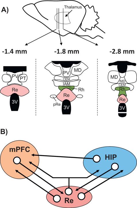
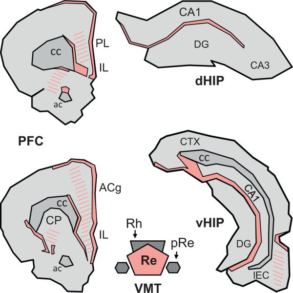
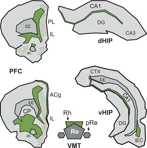
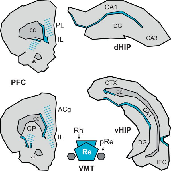
The reuniens and rhomboid nuclei, located in the ventral midline of the thalamus, have long been regarded as having non-specific effects on the cortex, while other evidence suggests that they influence behavior related to the photoperiod, hunger, stress or anxiety. We summarise the recent anatomical, electrophysiological and behavioral evidence that these nuclei also influence cognitive processes. The first part of this review describes the reciprocal connections of the reuniens and rhomboid nuclei with the medial prefrontal cortex and the hippocampus. The connectivity pattern among these structures is consistent with the idea that these ventral midline nuclei represent a nodal hub to influence prefrontal-hippocampal interactions. The second part describes the effects of a stimulation or blockade of the ventral midline thalamus on cortical and hippocampal electrophysiological activity. The final part summarizes recent literature supporting the emerging view that the reuniens and rhomboid nuclei may contribute to learning, memory consolidation and behavioral flexibility, in addition to general behavior and aspects of metabolism.

Keywords: 5-CSRT; 5-choice serial reaction time; Alv; Behavioral flexibility; CRF; DNMTP; EEG; FG; FR; GABA; Hippocampus; IL; LTP; Lac-mol; MFB; MdT; Medial prefrontal cortex; N-methyl-D-aspartate; N-methyl-D-aspartate receptor; NGF; NMDA; NMDAR; Nerve growth factor; Non specific thalamus; Ori; PHA-L; PL; Phaseolus vulgaris leucoagglutinin; Re; Reference memory; Reuniens nucleus; Rh; Rhomboid nucleus; Spatial memory; Systems-level consolidation; Ventral midline thalamus; Working memory; alveus; corticotrophin releasing factor; delayed nonmatching to place (or to position); electroencephalogram or electroencephalographic; fluorogold; fluororuby; gamma aminobutyric acid; infralimbic cortex; long term potentiation; mPFC; medial forebrain bundle; medial prefrontal cortex; midline thalamus; mol; pRe; perireuniens nucleus; prelimbic cortex; pyr; rad; reuniens nucleus; rhomboid nucleus; sEPSCs; spontaneous excitatory post-synaptic currents; stratum lacunosum-moleculare; stratum moleculare; stratum oriens; stratum pyramidale; stratum radiatum.


Figures

Fig. 1
Fig. 1
Neuroanatomical organization of the midline thalamus (A) and connectivity diagram between the prefrontal cortex, the reuniens nucleus and the hippocampus (B). A: Nuclei of the ventral midline thalamus, with particular focus on the reuniens (Re) and rhomboid (Rh) nuclei at 3 anterior-posterior levels (the most caudal is on the left). Abbreviations: CEM, central medial nucleus; IAM, interanteromedial nucleus; IMD, interomediodorsal nucleus; MD, mediodorsal nucleus; pRe, perireuniens nuclei; PT, paratenial nucleus; PV, paraventricular nucleus; Re: reuniens nucleus; Rh, rhomboid nucleus. B: Schematic representation of the organization of the network connectivity within a system formed by the reuniens nucleus (Re), which is the largest of the ventral midline thalamus, the hippocampus (HIP) and the medial prefrontal cortex (mPFC). It is noteworthy that whereas the HIP has pronounced projections to the mPFC, there are no direct return ones from the mPFC to the hippocampus. The Re has dense projections to both the mPFC and the HIP (see also Fig. 2), and a small proportion of neurons of the Re (between 3 and 6%) even send axon collaterals to both structures, as described very recently (Hoover and Vertes, 2012). Finally, the mPFC and the HIP have dense projections to the Re/Rh. This organization places the Re, and perhaps more generally the ventral midline thalamus (which also encompasses the rhomboid and perireuniens nuclei), in a pivotal position to influence prefrontal cortical and hippocampal functions, and perhaps even in a more specific way functions which depend on information exchange between or coordination of these two structures.
Fig. 2
Fig. 2
Terminal projection fields of the reuniens nucleus in the prefrontal cortex and the hippocampus of the Rat. Illustration of the location and extent of the regions of densest (filled) and weaker (hatched) fiber staining at two A-P levels of the medial prefrontal cortex (mPFC) and in the dorsal and ventral hippocampus (dHIP and vHIP, respectively) produced by an injection of the anterograde anatomical tracer Phaseolus vulgaris leucoagglutinin into the Re. The drawings were made according to the darkfield microphotographs (Figs. 5 and 6) and the schematic representations (Fig. 3) shown in Vertes et al. (2006). Abbreviations: ac, anterior commissure; ACg, anterior cingulate cortex; cc, corpus callosum; CP, caudate putamen; DG, dentate gyrus; IL, infralimbic cortex; LEC, lateral entorhinal cortex; pRe, perireuniens nucleus; PL, prelimbic cortex; Re, reuniens nucleus; Rh, rhomboid nucleus.
Fig. 3
Fig. 3
Terminal projection fields of the rhomboid nucleus in the prefrontal cortex and the hippocampus of the Rat. Illustration of the location and extent of the regions of densest (filled) and of weaker (hatched) fiber staining at two A-P levels of the medial prefrontal cortex (mPFC) and in the dorsal and ventral hippocampus (dHIP and vHIP, respectively) produced by an injection of the anterograde anatomical tracer Phaseolus vulgaris leuccoagglutinin into the Rh. The drawings were made according to the darkfield microphotographs (Figs. 9–12) and the schematic representations (Fig. 9) shown in Vertes et al. (2006). Abbreviations: ac, anterior commissure; ACg, anterior cingulate cortex; cc, corpus callosum; CP, caudate putamen; DG, dentate gyrus; IL, infralimbic cortex; LEC, lateral entorhinal cortex; pRe, perireuniens nucleus; PL, prelimbic cortex; Re, reuniens nucleus; Rh, rhomboid nucleus.
Fig. 4
Fig. 4
Regions in which the terminal projection fields of the reuniens nucleus and the rhomboid nucleus show a clear-cut overlapping. The filled regions delimited by continuous lines correspond to the areas in which densest staining after PHA-L injections into the Re overlaps with densest staining after injection of the same tracer into the Rh. The hatched areas correspond to the region where a weaker but nonetheless clear-cut density of projections from one nucleus overlaps with a strong or a weaker density of projections from the other nucleus. This figure has been drawn from Figs. 2 and 3 of the current article.
Fig. 5
Fig. 5
Functional scheme of the connectivity between the reuniens nucleus and the hippocampus. Drawing providing a summary of the main findings reported by Dolleman-van der Weel et al. (1997) and hence derived connectivity loop which they proposed. Neurons from the caudal region of the Re project to the rostral Re, from where neurons establish monosynaptic contacts with the dendrites of pyramidal CA1 cells in the stratum lacunosum-moleculare, of interneurons with their soma in the stratum radiatum, and of interneurons with their soma in the stratum oriens. Most contacts in this model are excitatory, except the contacts of the interneurons of the stratum oriens which mediate feedforward inhibition on CA1 pyramidal neurons. The axons of the latter, which course in the alveus, project back to the Re via the subiculum. A stimulation of the Re using a paired stimulation protocol will produce facilitation and exhibit two types of evoked activity profiles in the stratum lacunosum-moleculare depicted as simple (one negative deflection/stimulation) and complex (2 deflections/stimulation). The complex profiles are obtained with caudal stimulation of Re and most probably correspond to the disynaptic EPSPs. This figure has been drawn after Fig. 6 in Dolleman-van der Weel et al. (1997). Abbreviations: alv: alveus; lac-mol: stratum lacunosum-moleculare; ori: stratum oriens; pyr: stratum pyramidale; rad: stratum radiatum.
Fig. 6
Fig. 6
The 5-choice serial reaction time task. This test measures sustained visual attention in rodents. It uses a sound-attenuated chamber with five light stimulus presentation modules on one rear wall. The chamber is illuminated by a house light (yellow bulb + stripes), which can be turned off upon request (e.g., in order to indicate a failed trial). Each of the five modules is equipped with an infrared photocell beam enabling both light stimulus presentation (e.g., as indicated by the red triangle in B) and detection of a rat's nose-poke in the hole of the module. At the opposite, a reward pellet is delivered from an automated food magazine, but only when the rat has made a correct response. At the start of the test, the rat faces the 5 light-presentation modules (A), focusing visual attention on them (as symbolized by the white triangle). A brief light signal (0.5 s or even less; red triangle) is presented in one of the modules (B). Upon detection, the rat has to move towards this module and make a nose-poke in its hole (C). If so, the trial is recorded as correct and a reward pellet is delivered automatically at the opposite wall from an engine-driven magazine (D). A nose-poke in any other module is counted as an incorrect trial. No nose-poke before a fixed delay is counted as an omission. A nose-poke during the hold period preceding the light signal in the module is considered a premature response. Repeated nose-pokes in the same module despite a light signal that has been shifted to another module are accounting for perseverative responses. In case of a correct response, once the pellet has been collected, the next trial is started after a fixed (light signal occurrence can be anticipated by the rat) or a variable (light signal occurrence cannot be anticipated by the rat) hold period.
Fig. 7
Fig. 7
A role for the reuniens nucleus and the rhomboid nucleus in systems-level consolidation of a memory. (A) During encoding, the different perceptual features of an experience are processed in primary and associative areas of the cortex, from where a representation is integrated in the hippocampus as a recent memory trace. (B) Over time, off line reactivation – probably during particular stages of sleep – of hippocampo-cortical networks progressively leads to a strengthening of existing connections within and between cortical modules as well as to the establishment of new connections therein. This process probably requires bidirectional information flow between the mPFC and the hippocampus, wherein the Re and Rh nuclei might play the role of a hub relaying at least the information transmission from the prefrontal cortex to the hippocampus. (C) These strengthened connections in the cortex provide, in whole or in part, cortical support to remote memories, which therefore may become, though not necessarily completely as shown by e.g., Lopez et al. (2012), independent of the hippocampus. This figure has been drawn after Frankland and Bontempi (2005).
Fig. 8
Fig. 8
The Re/Rh lesion prevents the formation of a remote memory for place. (A) Photograph of the water maze in which the rats were trained and tested. (B) Location of the platform (white circle) in the water maze during the 8-day training protocol (4 trials/day). (C) Typical swim paths as recorded during a probe trial given 5 or 25 days after the end of training in rats subjected to Re/Rh lesions before training. (D) Time spent in the target quadrant (former location of the platform) during the probe trial 5 and 25 days post-acquisition in sham-operated control rats and rats with Re/Rh lesions (analysis of lesion location and extent showed no difference between rats tested at the delay of 5 post-acquisition days vs. those tested at that of 25 days). The * indicates a significant difference with lesion rats tested at the delay of 5 days (p < 0.001). This figure has been drawn after Loureiro et al. (2012).
Fig. 9
Fig. 9
The double-H maze. Photograph showing a general picture of the testing device as it was installed for our experiment assessing behavioral flexibility (see Fig. 10 for an illustrated summary of the protocol used). Holding in a square of 160 cm × 160 cm and placed on a 80-cm high table, this device has been filled with opaque water (obtained by addition of powdered milk) to about 14 cm height. For each training session, an invisible escape platform is immerged in the water at the extremity of a constant arm; it is removed for each of the two probe trials. For further details about this test and protocol variants, see e.g., Cassel et al. (2012) and Pol-Bodetto et al. (2011).
Fig. 10
Fig. 10
Analysis of strategy shifting capabilities in the double-H maze after reversible inactivation of the Re/Rh. (A) Time line of the experiment. After a habituation day (0), rats were trained for two consecutive days (1,2), given a first 24 h-delayed probe trial (3), trained for two additional days to strengthen learning (4,5) and given a second 24 h-delayed probe trial (6). On each training day they were released from the north (N) or south (S) arm according to the indicated sequence. Before each probe trial, rats were infused with muscimol (MSCI; 250 ng in 0.3 μL) or PBS. (B) Illustration of the configurations used during maze training. The rats were given 4 daily trials for which they were released twice from the S and twice from the N (in a randomized order; e.g., N,S,S,N, then S,N,N,S, then N,S,N,S. . .). The platform was always located in the NE arm. The arm opposite to the one in which the rats were placed at the start of a trial was always closed by a transparent guillotine door (it corresponds to the grey-filled arm). Rats could reach the target arm by one of both efficient response behavior (left-left or right-left turn sequences) or learn the place, and most probably, as shown earlier (Pol-Bodetto et al., 2011), made both in the place-response order. (C) On days 3 and 6, thirty min before each probe trial, about half the rats were infused with MSCI in the Re/Rh, the other half being infused with an equivalent volume of PBS as a control. For both probe trials, rats were released from the SW arm, the NW one was closed by a guillotine door; the platform had been removed from the device. We analyzed the capability of the rats to shift to a response based on place memory either immediately or after having entered the N arm. We found that in rats subjected to MSCI infusions into the Re/Rh such a shift was impossible, as also found in rats subjected to exactly the same protocol but given the infusions (250 ng in 1 μL) into either the mPFC or the hippocampus (Loureiro et al., unpublished data). This figure has been adapted from Cholvin et al. (2013).

References

    1. Abela AR, Dougherty SD, Fagen ED, Hill CJ, Chudasama Y. Inhibitory control deficits in rats with ventral hippocampal lesions. Cereb. Cortex. 2013;23(6):1396–1409. - PubMed
    1. Aggleton JP, Brown MW. Episodic memory, amnesia, and the hippocampal-anterior thalamic axis. Behav. Brain Sci. 1999;22:425–444. - PubMed
    1. Aggleton JP, O'Mara SM, Vann SD, Wright NF, Tsanov M, Erichsen JT. Hippocampal-anterior thalamic pathways for memory: uncovering a network of direct and indirect actions. Eur. J. Neurosci. 2010;31(12):2292–2307. - PMC - PubMed
    1. Aggleton JP, Dumont JR, Warburton EC. Unraveling the contributions of the diencephalon to recognition memory: a review. Learn. Mem. 2011;18(6):384–400. - PMC - PubMed
    1. Amici S. Thalamic infarcts and haemorrhages. Front. Neurol. Neurosci. 2012;30:132–136. - PubMed

Publication types

MeSH terms