. 2013 Sep 19;501(7467):380-4.
doi: 10.1038/nature12530. Epub 2013 Sep 11.

Usp16 contributes to somatic stem-cell defects in Down's syndrome

Affiliations

Usp16 contributes to somatic stem-cell defects in Down's syndrome

Maddalena Adorno et al. Nature. .

Abstract

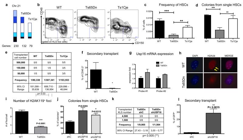
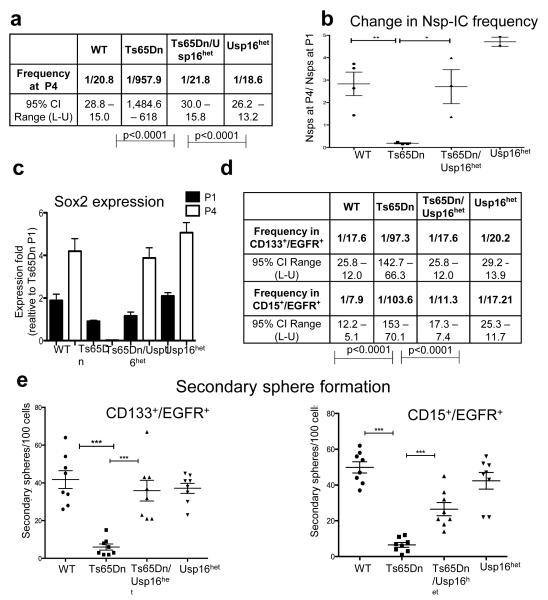
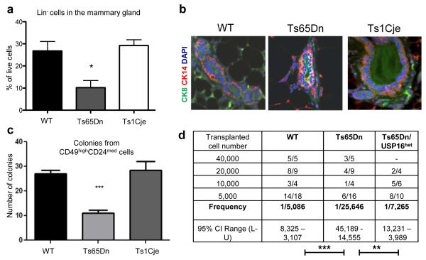
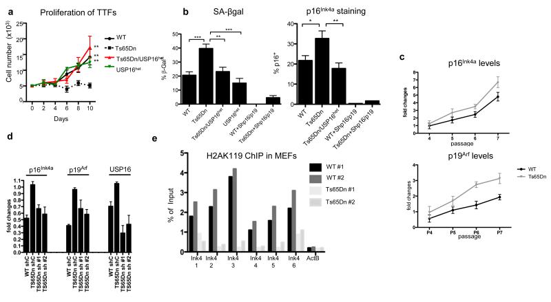
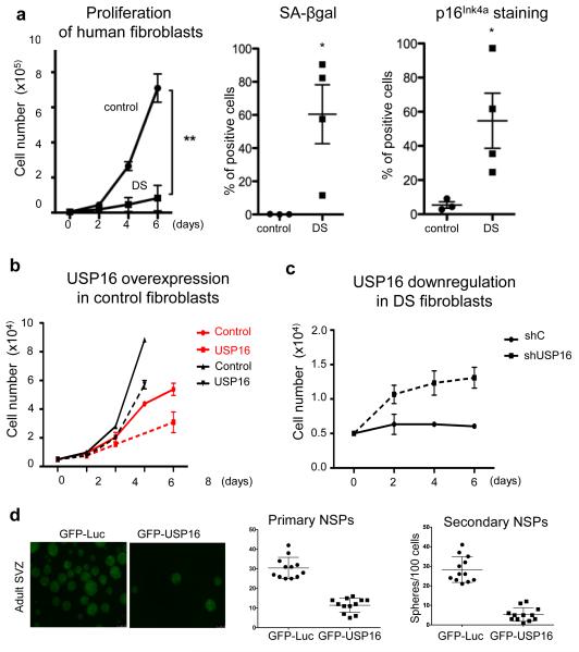
Down's syndrome results from full or partial trisomy of chromosome 21. However, the consequences of the underlying gene-dosage imbalance on adult tissues remain poorly understood. Here we show that in Ts65Dn mice, which are trisomic for 132 genes homologous to genes on human chromosome 21, triplication of Usp16 reduces the self-renewal of haematopoietic stem cells and the expansion of mammary epithelial cells, neural progenitors and fibroblasts. In addition, Usp16 is associated with decreased ubiquitination of Cdkn2a and accelerated senescence in Ts65Dn fibroblasts. Usp16 can remove ubiquitin from histone H2A on lysine 119, a critical mark for the maintenance of multiple somatic tissues. Downregulation of Usp16, either by mutation of a single normal Usp16 allele or by short interfering RNAs, largely rescues all of these defects. Furthermore, in human tissues overexpression of USP16 reduces the expansion of normal fibroblasts and postnatal neural progenitors, whereas downregulation of USP16 partially rescues the proliferation defects of Down's syndrome fibroblasts. Taken together, these results suggest that USP16 has an important role in antagonizing the self-renewal and/or senescence pathways in Down's syndrome and could serve as an attractive target to ameliorate some of the associated pathologies.


Figures

Figure 1
Figure 1. Usp16 contributes to defective HSCs in Ts65Dn mice
a, DS mouse models. b,c, HSC frequency is decreased in Ts65Dn, but not in Ts1Cje bone marrow. Representative plots are shown (n=4). d, Colony formation assay from single HSCs. (n=3). e, Limiting dilution analysis (ELDA) of marrow cells. f, Secondary engraftment three months after transplantation, shown as percentage of donor cells in the peripheral blood. Transplants were repeated twice. g, Quantification of Usp16 mRNA. h,i, H2AK119 immunofluorescence in HSCs. The arrows indicate the stained H2AK119 foci in one representative picture. Bottom panel, quantification of positive foci (n=100 cells analyzed in two experiments). j, Knockdown of Usp16 in Ts65Dn HSCs rescues colony formation (n=3). k,l, Knockdown of Usp16 improves the engraftment potential of Ts65Dn and their ability to undergo secondary transplantation. Transplants were repeated twice.
Figure 2
Figure 2. Ts65Dn mice, but not Ts65Dn/Usp16het mice, show defective neuralprogenitor cells
a, ELDA for SVZ Lin cells (at P4). b,c, Nsp-IC frequency and Sox2 mRNA levels decrease only in Ts65Dn culture. d,e, ELDA and secondary sphere formation experiments with the indicated populations show an neural progenitor expansion deficit specific to Ts65Dn mice. (n=3 per group in all experiments).
Figure 3
Figure 3. Ts65Dn mammary cells are affected by levels of Usp16
a, Lin cells are reduced in Ts65Dn, but not in Ts1Cje glands. (n=5). b, Immunofluorescence staining shows an increase overlapping of luminal and basal cytokeratins in Ts65Dn glands. c, Ts65Dn sorted MRUs form fewer colonies then controls (n=4) d, ELDA shows a decrease number of repopulating cells in Ts65Dn, but not in Ts65Dn/Usp16het glands. Three independent experiments were performed, two for Ts65Dn/Usp16het.
Figure 4
Figure 4. Usp16 contributes to proliferation defects and senescence in Ts65Dn fibroblasts
a, Proliferation of TTFs seeded at P2. b, Senescent cells are frequent in Ts65Dn, but not in Ts65Dn/USP16het TTFs, as shown by SA-βgal and p16Ink4a staining at P3. Downregulation of cdkn2a blocks senescence. c, p16Ink4a and p19Arf mRNA levels rapidly increase during passaging in Ts65Dn MEFs. d, Downregulation of Usp16 normalizes mRNA expression of Ink4a/Arf by P6 Ts65Dn MEFs. e, ChIP analyses at the cdkn2a locus show lower levels of H2AK119 binding by two Ts65Dn MEF chromatin samples.
Figure 5
Figure 5. Human cells are affected by USP16 levels
a, Proliferation analysis, as well as SA-βgal and p16Ink4a staining, of three control and four DS human fibroblast cultures show growth impairment and senescence of DS cells. b,c, Lentiviral overexpression of USP16 decreases the proliferation of two different control fibroblast lines, while donwregulation of USP16 in DS fibroblasts promotes proliferation. d, Overexpression of USP16 reduces the formation of neurospheres derived from human adult SVZ cells. The right panel quantifies the number of spheres in the first and second passage. (p<0.0001). All the experiments were replicated at least twice.

Comment in

References

    1. Antonarakis SE, Lyle R, Dermitzakis ET, Reymond A, Deutsch S. Chromosome 21 and down syndrome: from genomics to pathophysiology. Nat. Rev. Genet. 2004;5:725–738. - PubMed
    1. Yang Q, Rasmussen SA, Friedman JM. Mortality associated with Down’s syndrome in the USA from 1983 to 1997: a population-based study. Lancet. 2002;359:1019–1025. - PubMed
    1. Roth GM, Sun B, Greensite FS, Lott IT, Dietrich RB. Premature aging in persons with Down syndrome: MR findings. AJNR Am J Neuroradiol. 1996;17:1283–1289. - PMC - PubMed
    1. Zigman WB, Lott IT. Alzheimer’s disease in Down syndrome: neurobiology and risk. Ment Retard Dev Disabil Res Rev. 2007;13:237–246. - PubMed
    1. Liu L, Rando TA. Manifestations and mechanisms of stem cell aging. J. Cell Biol. 2011;193:257–266. - PMC - PubMed

Publication types

MeSH terms