. 2013 Dec 15;305(12):C1230-9.
doi: 10.1152/ajpcell.00053.2013. Epub 2013 Sep 11.

Histone deacetylase 6-mediated deacetylation of α-tubulin coordinates cytoskeletal and signaling events during platelet activation

Affiliations

Histone deacetylase 6-mediated deacetylation of α-tubulin coordinates cytoskeletal and signaling events during platelet activation

Joseph E Aslan et al. Am J Physiol Cell Physiol. .

Abstract

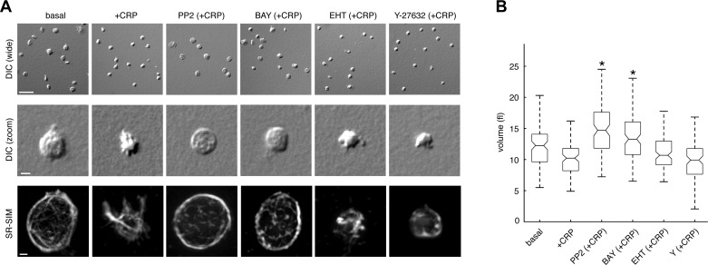
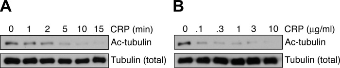
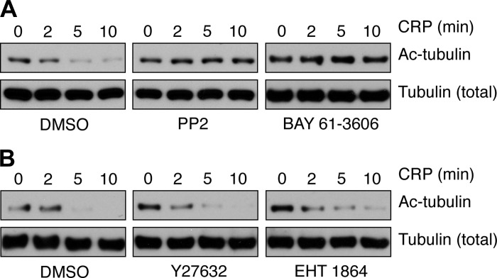
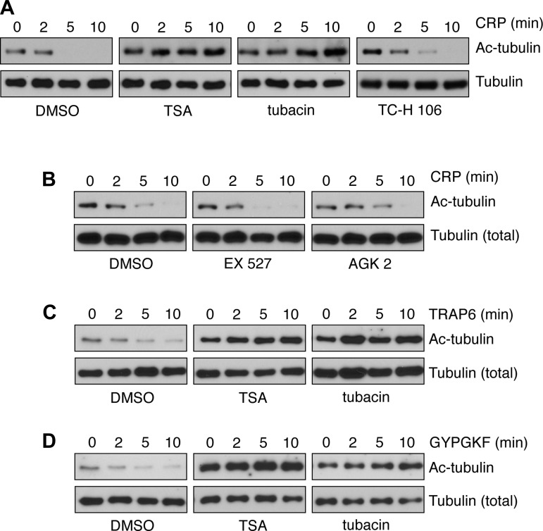
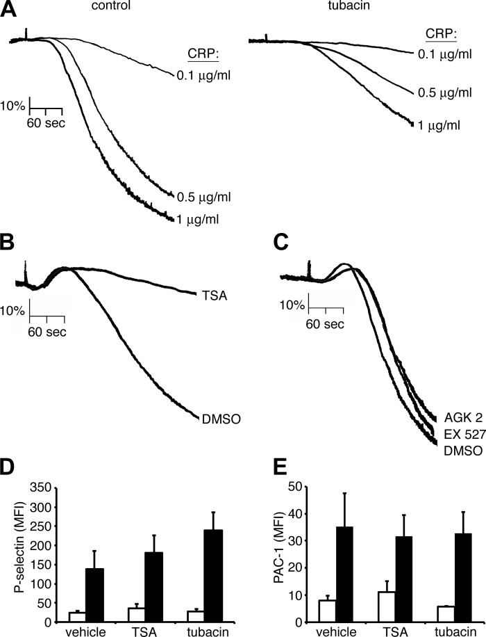
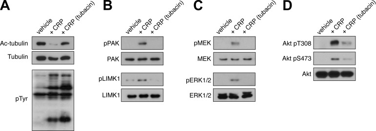
The tubulin cytoskeleton plays a key role in maintaining the characteristic quiescent discoid shape of resting platelets. Upon activation, platelets undergo a dramatic change in shape; however, little is known of how the microtubule system contributes to regulating platelet shape and function. Here we investigated the role of the covalent modification of α-tubulin by acetylation in the regulation of platelet physiology during activation. Superresolution microscopy analysis of the platelet tubulin cytoskeleton showed that the marginal band together with an interconnected web of finer tubulin structures collapsed upon platelet activation with the glycoprotein VI (GPVI)-agonist collagen-related peptide (CRP). Western blot analysis revealed that α-tubulin was acetylated in resting platelets and deacetylated during platelet activation. Tubacin, a specific inhibitor of the tubulin deacetylase HDAC6, prevented tubulin deacetylation upon platelet activation with CRP. Inhibition of HDAC6 upregulated tubulin acetylation and disrupted the organization of the platelet microtubule marginal band without significantly affecting platelet volume changes in response to CRP stimulation. HDAC6 inhibitors also inhibited platelet aggregation in response to CRP and blocked platelet signaling events upstream of platelet Rho GTPase activation. Together, these findings support a role for acetylation signaling in controlling the resting structure of the platelet tubulin marginal band as well as in the coordination of signaling systems that drive platelet cytoskeletal changes and aggregation.

Keywords: HDAC6; acetylation; platelets; tubulin.


Figures

Fig. 1.
Fig. 1.
Platelet activation results in a decrease in platelet volume and condensation of the tubulin cytoskeleton. A: replicate samples of purified human platelets were treated with the glycoprotein VI (GPVI)-agonist agonist collagen-related peptide (CRP; 1 μg/ml) in solution for 5 min in the presence of vehicle or the Src family kinase (SFK) inhibitor PP2 (10 μM), the Syk inhibitor BAY 61-3606 (BAY; 10 μM), the Rac inhibitor EHT 1864 (EHT; 50 μM), or the Rho kinase inhibitor Y-27632 (Y; 10 μM). Platelets were then fixed and visualized by differential interference contrast (DIC) microscopy. Wide field scale bar, 10 μm. Zoom field scale bar, 2 μm. Tubulin fluorescence of representative platelets was visualized by superresolution structured illumination microscopy (SR-SIM) and reconstructed into 3 dimensions using Imaris software. Scale bar, 500 nm. B: box plots of platelet volume determined by DIC optical sectioning microscopy as described in materials and methods. Central box marks indicate median values of platelet volumes; box edges represent 25th and 75th percentile values. Whiskers extend to the most extreme nonoutlier data points within ±2.7σ, where σ is the standard deviation of the data. *Inhibitor-treated samples with volumes significantly greater than that of vehicle-treated samples in the presence of CRP.
Fig. 2.
Fig. 2.
Time- and concentration-dependent deacetylation of tubulin upon platelet activation by CRP. A: purified human platelets were treated with CRP in solution (1 μg/ml) for 1–15 min before lysis into sample buffer and Western blot analysis for acetylated α-tubulin (Ac-tubulin) Lys 40. B: purified human platelets were treated with CRP (0.1–10 μg/ml) for 5 min before lysis into sample buffer and Western blot analysis. Total tubulin levels are shown as a control for protein loading. Results are representative of three separate experiments.
Fig. 3.
Fig. 3.
Platelet signaling inhibition blocks CRP-triggered tubulin deacetylation. Replicate samples of purified human platelets were pretreated with the SFK inhibitor PP2 (10 μM), the Syk inhibitor BAY 61-3606 (10 μM) (A), the Rho kinase (ROCK) inhibitor Y-27632 (10 μM), the Rac inhibitor EHT 1864 (50 μM) (B), or vehicle alone before stimulation with 1 μg/ml CRP (2–10 min). Platelets were lysed into sample buffer and analyzed for tubulin acetylation by Western blotting. Results are representative of three separate experiments.
Fig. 4.
Fig. 4.
Histone deacetylase (HDAC) protein expression and activity in human platelets. A: purified human platelets and MDA-MB-231 cells were lysed directly into sample buffer and analyzed for HDAC 1, 2, 4, 6, and 7 and sirtuin 2 (SIRT2) protein expression by Western blotting. Tubulin serves as a loading control for total cellular protein. B: human platelets were treated with the pan-HDAC inhibitor trichostatin A (TSA; 10 μM), the HDAC6-specific inhibitor tubacin (10 μM), or vehicle alone before lysis into assay buffer and analysis of total HDAC activity (means ± SE; n = 4). *P < 0.05 compared with vehicle-treated platelets. C and D: replicate preparations of purified human platelets (n = 3) were fixed, immobilized on coverglass, stained for the presence of HDAC6 and α-tubulin protein, and examined by fluorescence deconvolution microscopy (C) or stained with α-tubulin and acetylated tubulin and visualized by SR-SIM (D). Scale bar, 1 μm.
Fig. 5.
Fig. 5.
HDAC6 inhibition prevents platelet tubulin deacetylation. A and B: purified human platelets were pretreated with the pan-HDAC inhibitor TSA (10 μM), the HDAC6-specific inhibitor tubacin (10 μM), the HDAC1/3 inhibitor TC-H 106 (10 μM), or vehicle alone (DMSO) (A) or the SIRT1 inhibitor EX 527 (10 μM), the SIRT2 inhibitor AGK2 (10 μM), or vehicle alone (DMSO) (B) before stimulation with 1 μg/ml CRP (2–10 min). Platelets were lysed into sample buffer and analyzed for tubulin acetylation by Western blot. C and D: purified human platelets were pretreated with the pan-HDAC inhibitor TSA (10 μM), the HDAC6-specific inhibitor tubacin (10 μM), or vehicle alone (DMSO) before stimulation (2–10 min) with the PAR1 agonist peptide TRAP6 (C) or the PAR4 agonist peptide GYPGKF (D). Western blot results are each representative of 4 experiments.
Fig. 6.
Fig. 6.
HDAC6 inhibition alters the structure of the platelet marginal band. A: purified human platelets were pretreated with TSA (10 μM), tubacin (10 μM), or vehicle alone before stimulation with 1 μg/ml CRP. After 5 min, platelets were fixed, immobilized on coverglass, stained with antibodies directed against α-tubulin, and examined by SR-SIM. Representative 0.56-μm slices from three separate experiments are shown. Wide field scale bar, 10 μm. Zoomed image scale bar, 1 μm. BD: after fixation, platelets were also examined by DIC microscopy (B and C) and quantitative volumetric analysis (D) as described in materials and methods. Central box marks indicate median values of platelet volume; box edges represent 25th and 75th percentile values. Whiskers extend to the most extreme nonoutlier data points. *Values significantly lower than that of resting platelets (basal).
Fig. 7.
Fig. 7.
HDAC inhibition blocks platelet aggregation in solution. A: washed human platelets (2 × 10⁸/ml) were incubated with vehicle (DMSO) or the HDAC6-specific inhibitor tubacin (10 μM) prior to stimulation with increasing concentrations of CRP, and the change in optical density indicative of platelet aggregation was recorded. Aggregation traces representative of three separate experiments are shown. B and C: washed human platelets (2 × 10⁸/ml) were incubated with vehicle (DMSO) or the pan-HDAC inhibitor TSA (10 μM) (B) or the SIRT1 inhibitor EX 527 (10 μM) or the SIRT2 inhibitor AGK2 (10 μM) (C) prior to stimulation with CRP (10 μg/ml) in the presence of 2 U/ml apyrase, and the change in optical density indicative of platelet aggregation was recorded. D and E: platelet surface P-selectin levels (D) and integrin αIIbβ3 activation (PAC-1) (E) analyzed by flow cytometry following vehicle (DMSO), TSA, and tubacin treatment and incubation with vehicle (white bars) or CRP (1 μg/ml; black bars) stimulation. MFI, mean fluorescence intensity. Data are represented as means ± SE.
Fig. 8.
Fig. 8.
HDAC6 mediates p21-activated kinase (PAK), MAPK, and Akt signaling in platelets. Purified human platelets were pretreated with tubacin (10 μM) or vehicle prior to stimulation with CRP (1 μg/ml). After 5 min, platelets were lysed in sample buffer and analyzed by Western blotting for acetyl tubulin, total phosphotyrosine (pTyr) moieties (A), phosphorylated PAK Ser 141, phosphorylated LIM domain kinase (LIMK1) Thr 508 (B), phospho-MEK, phospho-ERK (C), and phospho-Akt Thr 308 and Ser 473 (D). Western blotting results are representative of six separate experiments.

Comment in

References

    1. Arias-Romero LE, Chernoff J. A tale of two Paks. Biol Cell 100: 97–108, 2008 - PubMed
    1. Aslan JE, Baker SM, Loren CP, Haley KM, Itakura A, Pang J, Greenberg DL, David LL, Manser E, Chernoff J, McCarty OJ. The PAK system links Rho GTPase signaling to thrombin-mediated platelet activation. Am J Physiol Cell Physiol 305: C519–C528, 2013 - PMC - PubMed
    1. Aslan JE, Itakura A, Gertz JM, McCarty OJ. Platelet shape change and spreading. Methods Mol Biol 788: 91–100, 2012 - PubMed
    1. Aslan JE, Itakura A, Haley KM, Tormoen GW, Loren CP, Baker SM, Pang J, Chernoff J, McCarty OJ. p21 activated kinase signaling coordinates glycoprotein receptor VI-mediated platelet aggregation, lamellipodia formation, and aggregate stability under shear. Arterioscler Thromb Vasc Biol 33: 1544–1551, 2013 - PMC - PubMed
    1. Aslan JE, McCarty OJ. Rho GTPases in platelet function. J Thromb Haemost 11: 35–46, 2013 - PMC - PubMed

Publication types

MeSH terms

LinkOut - more resources