. 2013 Oct;140(20):4203-13.
doi: 10.1242/dev.095562. Epub 2013 Sep 11.

Nkx genes are essential for maintenance of ventricular identity

Affiliations

Nkx genes are essential for maintenance of ventricular identity

Kimara L Targoff et al. Development. 2013 Oct.

Abstract

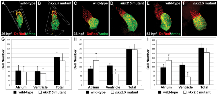
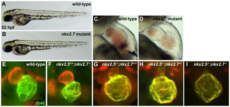
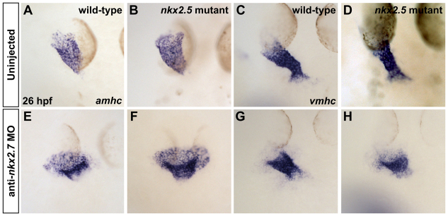
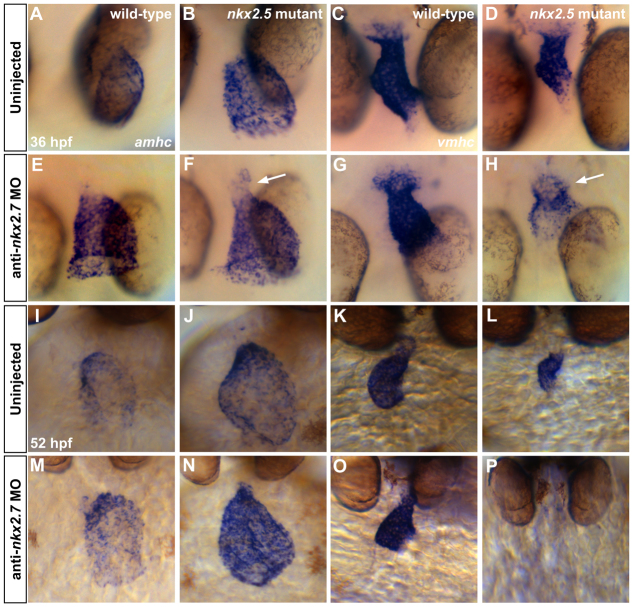
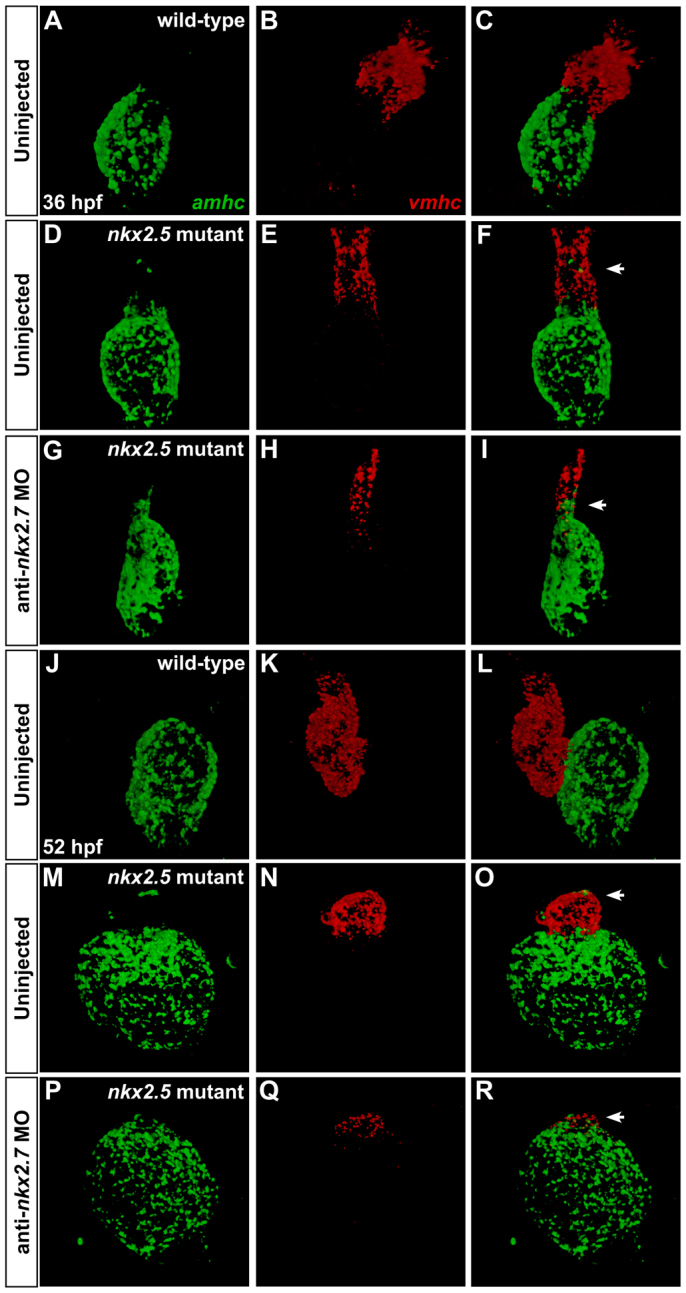
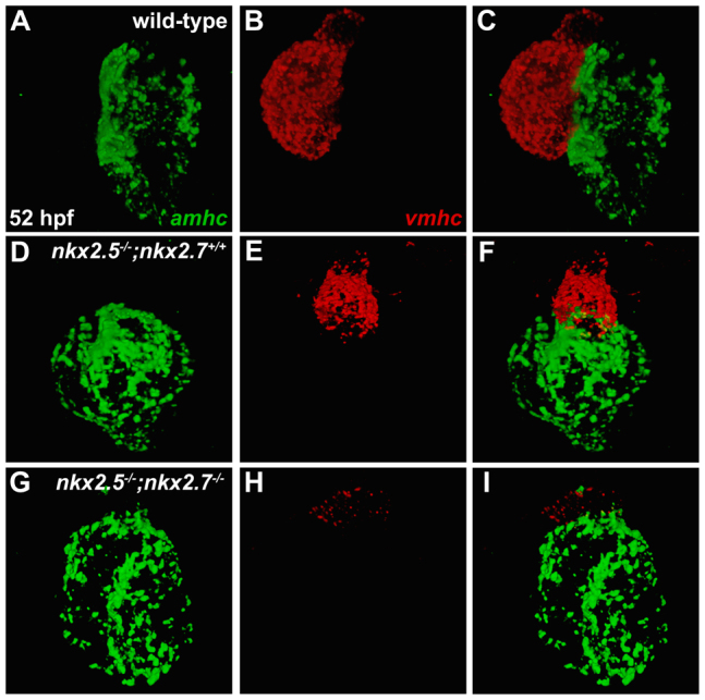
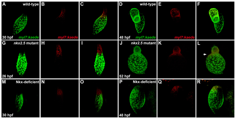
Establishment of specific characteristics of each embryonic cardiac chamber is crucial for development of a fully functional adult heart. Despite the importance of defining and maintaining unique features in ventricular and atrial cardiomyocytes, the regulatory mechanisms guiding these processes are poorly understood. Here, we show that the homeodomain transcription factors Nkx2.5 and Nkx2.7 are necessary to sustain ventricular chamber attributes through repression of atrial chamber identity. Mutation of nkx2.5 in zebrafish yields embryos with diminutive ventricular and bulbous atrial chambers. These chamber deformities emerge gradually during development, with a severe collapse in the number of ventricular cardiomyocytes and an accumulation of excess atrial cardiomyocytes as the heart matures. Removal of nkx2.7 function from nkx2.5 mutants exacerbates the loss of ventricular cells and the gain of atrial cells. Moreover, in these Nkx-deficient embryos, expression of vmhc, a ventricular gene, fades, whereas expression of amhc, an atrial gene, expands. Cell-labeling experiments suggest that ventricular cardiomyocytes can transform into atrial cardiomyocytes in the absence of Nkx gene function. Through suggestion of transdifferentiation from ventricular to atrial fate, our data reveal a pivotal role for Nkx genes in maintaining ventricular identity and highlight remarkable plasticity in differentiated myocardium. Thus, our results are relevant to the etiologies of fetal and neonatal cardiac pathology and could direct future innovations in cardiac regenerative medicine.

Keywords: Atrium; Chamber identity; Ventricle; Zebrafish; nkx2.5; nkx2.7.


Figures

Fig. 1.
Fig. 1.
Mutation of nkx2.5 disrupts cardiac morphogenesis. (A,B) Lateral views of live zebrafish embryos, anterior to the left, at 52 hpf. Other than cardiac defects and pericardial edema, the nkx2.5 mutant embryo (B) appears morphologically normal. (C,D) Lateral views of live embryos, anterior to the top, at 52 hpf. In contrast to the wild-type heart (C), the nkx2.5 mutant heart (D) is unlooped and has striking defects in both ventricular and atrial morphology. (E,F) MF20/S46 immunofluorescence distinguishes ventricular myocardium (red) from atrial myocardium (yellow). Frontal views, anterior to the top, at 52 hpf. In comparison to the wild-type heart (E), the nkx2.5 mutant heart (F) has a diminutive ventricle and an enlarged atrium. (G-L) In situ hybridization depicts expression of myl7 (G,H), vmhc (I,J) and amhc (K,L) in wild-type (G,I,K) and nkx2.5 mutant (H,J,L) embryos. Dorsal views, anterior to the top, at 26 hpf. (G,H) nkx2.5 mutant embryos exhibit subtle defects in heart tube extension, including a broad inflow region and a compact outflow region. In nkx2.5 mutants, the ventricular portion of the heart tube is abnormally short and wide (I,J) and the atrial portion of the heart tube has a splayed appearance (K,L).
Fig. 2.
Fig. 2.
Increased atrial and decreased ventricular cell numbers are first evident in nkx2.5 mutants following heart tube extension. (A-F) Immunofluorescence indicates expression of the transgene Tg(-5.1myl7:nDsRed2) (red) in both cardiac chambers facilitating cardiomyocyte counting at 26 hpf (A,B), 36 hpf (C,D) and 52 hpf (E,F). Atria are labeled with the anti-Amhc antibody S46 (green). (G-I) Bar graphs indicate numbers of atrial and ventricular cardiomyocyte nuclei, as well as the total number of cardiomyocytes; mean and s.e.m. of each data set are shown, and asterisks indicate statistically significant differences from wild type (P<0.001). (G) At 26 hpf, we find no statistically significant difference in cell numbers in wild-type (n=20) and nkx2.5 mutant (n=7) embryos. (H) At 36 hpf, comparison of wild-type (n=16) and nkx2.5 mutant (n=10) embryos reveals an increase in atrial cell number and a decrease in ventricular cell number in nkx2.5 mutants. (I) At 52 hpf, comparison of wild-type (n=16) and nkx2.5 mutant (n=11) embryos reveals an increase in atrial cell number and a decrease in ventricular cell number in nkx2.5 mutants.
Fig. 3.
Fig. 3.
Loss of nkx2.7 function in nkx2.5 mutants yields a more prominent ventricular deficiency. (A-F) Frontal views, anterior to the top, of MF20/S46 immunofluorescence, which distinguishes ventricular myocardium (red) from atrial myocardium (yellow), at 52 hpf. In comparison to wild type (A), injection of anti-nkx2.7 MO (D) causes subtle abnormalities in chamber morphology. (B,E) In the nkx2.5 heterozygote, mild atrial enlargement is observed following injection of anti-nkx2.7 MO. (C,F) Injection of anti-nkx2.7 MO into nkx2.5 mutants yields severe loss of ventricular tissue and dramatic expansion of Amhc (S46, yellow) throughout the heart. Arrow indicates ventricular remnant that includes Amhc localization.
Fig. 4.
Fig. 4.
nkx2.5-/-;nkx2.7-/- double mutant demonstrates the synergistic requirement for Nkx genes in maintenance of ventricular identity. (A,B) Lateral views of live embryos, anterior to the left, at 52 hpf. General embryonic morphology appears similar in wild-type (A) and nkx2.7 mutant (B) embryos. (C,D) Lateral views of live embryos, anterior to the top, at 52 hpf. The nkx2.7 mutant heart (D) exhibits subtle defects in ventricular and atrial morphology. (E-I) Frontal views, anterior to the top, of MF20/S46 immunofluorescence, which distinguishes ventricular myocardium (red) from atrial myocardium (yellow), at 52 hpf. In comparison to wild-type embryos (E), nkx2.7 mutants (F) exhibit subtle abnormalities in chamber size and shape whereas nkx2.5 mutants (G) demonstrate an impressive size discrepancy between the ventricular and atrial chambers. (H) In the nkx2.5 mutant, loss of a single allele of nkx2.7 leads to increased localization of Amhc (S46, yellow) within the ventricle. (I) Exacerbation of the ventricular deficiency is evident in the nkx2.5-/-;nkx2.7-/- mutants, and Amhc is present throughout the entire cardiac structure.
Fig. 5.
Fig. 5.
Loss of nkx2.7 function in nkx2.5 mutants exacerbates defects during heart tube extension. (A-H) In situ hybridization depicts expression of amhc (A,B,E,F) and vmhc (C,D,G,H) in wild-type embryos (A,C), nkx2.5 mutants (B,D), wild-type embryos injected with anti-nkx2.7 MO (E,G) and Nkx-deficient embryos (F,H). Dorsal views, anterior to the top, at 26 hpf. The nkx2.5 mutant embryos exhibit subtle defects in heart tube extension, including a broader atrial region (A,B) and a slightly shorter ventricular region (C,D). Following MO injection, wild-type embryos demonstrate a spread of atrial cells and a compact coalescence of the ventricular cells (E,G). MO injection into nkx2.5 mutants leads to an exacerbated phenotype with a sprawling, widened atrial portion and a stunted ventricular portion of the heart tube (F,H).
Fig. 6.
Fig. 6.
Gradual elimination of ventricular gene expression and expansion of atrial gene expression in the Nkx-deficient heart. In situ hybridization illustrates expression of amhc (A,B,E,F,I,J,M,N) and vmhc (C,D,G,H,K,L,O,P) in wild-type embryos (A,C,I,K), nkx2.5 mutants (B,D,J,L), wild-type embryos injected with anti-nkx2.7 MO (E,G,M,O) and Nkx-deficient embryos (F,H,N,P). (A-H) Dorsal views, anterior to the bottom, at 36 hpf. The nkx2.5 mutants display a dilated atrium (A,B) and shortened ventricle (C,D). Following MO injection, wild-type embryos demonstrate a swollen atrium and widened ventricle (E,G). In nkx2.5 mutants, MO injection leads to distinct expansion of amhc and fading of vmhc in the ventricular remnant (F,H; arrows). (I-P) Ventral views, anterior to the top, at 52 hpf. Phenotypes are exacerbated at 52 hpf, both in terms of morphological defects and expression pattern changes. Most dramatically, in the Nkx-deficient embryo, amhc is expressed throughout the entire heart whereas vmhc expression is effectively abolished (N,P).
Fig. 7.
Fig. 7.
Dynamic expression patterns highlight gradual transitions in cardiomyocyte identity. Two-color fluorescence in situ hybridization facilitates sensitive detection of amhc (green) and vmhc (red) expression. Confocal projections of fixed, dissected wild-type (A-C,J-L), nkx2.5 mutant (D-F,M-O) and Nkx-deficient hearts (G-I,P-R); ventral views, anterior to the top. (A-I) At 36 hpf, ectopic amhc-expressing cells are found in the ventricle in the nkx2.5 mutant heart (D-F; arrowhead) and, more extensively, in the Nkx-deficient heart (G-I; arrowhead). (J-R) At 52 hpf, ectopic amhc-expressing cells are visualized in the dramatically diminished nkx2.5 mutant ventricle (M-O; arrowhead) whereas only a tuft of residual vmhc expression remains at the pole of the Nkx-deficient heart (P-R; arrowhead).
Fig. 8.
Fig. 8.
nkx2.5 and nkx2.7 work together to maintain vmhc expression and to limit amhc expression. (A-I) Fluorescence in situ hybridization depicts expression patterns of amhc (green) and vmhc (red) in wild-type (A-C), nkx2.5 mutant (D-F) and nkx2.5-/-;nkx2.7-/- double mutant (G-I) hearts at 52 hpf. Compared with robust expression of vmhc in both the wild-type (B) and nkx2.5 mutant (E) hearts, encroachment of ectopic amhc expression is demonstrated alongside dwindling vmhc expression in the ventricular remnant of the nkx2.5-/-;nkx2.7-/- mutant heart (G-I).
Fig. 9.
Fig. 9.
Ventricular-to-atrial transformation occurs in the absence of Nkx gene function. Confocal projections of live zebrafish hearts carrying Tg(myl7:kaede) are depicted immediately following photoconversion at 26-30 hpf (A-C,G-I,M-O) and after later visualization at 48-52 hpf (D-F,J-L,P-R); arterial pole to the top. Green fluorescence of Kaede (A,G,M) is converted to red fluorescence (B,H,N) following UV exposure of the region of interest; merged views (C,I,O) indicate that labeled cells are at the arterial pole of the heart tube. At later time points, retained red fluorescence (E,K,Q) is visualized together with the green Kaede (D,J,P) that is continually produced by the transgene; merged views (F,L,R) indicate the locations of the labeled cells. (A-F) In wild-type embryos, labeling of ventricular cells near the arterial pole of the heart tube (A-C) yields labeled cells within the ventricular chamber at 48 hpf (D-F) (n=17/17). (G-L) By contrast, comparable photoconversion of ventricular cardiomyocytes at the arterial pole of the nkx2.5 mutant heart tube (G-I) generates labeled cells both within the diminutive ventricular chamber and expanding into the atrial chamber (J-L, arrowhead in L) (n=10/12). (M-R) In Nkx-deficient embryos, labeled cells from the vmhc-expressing portion (supplementary material Fig. S9N) of the heart tube (M-O) are later detected within the single, amhc-expressing (supplementary material Fig. S9P) chamber of the heart (P-R) (n=2/2).

References

    1. Alexander J., Stainier D. Y., Yelon D. (1998). Screening mosaic F1 females for mutations affecting zebrafish heart induction and patterning. Dev. Genet. 22, 288–299 - PubMed
    1. Azpiazu N., Frasch M. (1993). tinman and bagpipe: two homeo box genes that determine cell fates in the dorsal mesoderm of Drosophila. Genes Dev. 7 7B, 1325–1340 - PubMed
    1. Bao Z. Z., Bruneau B. G., Seidman J. G., Seidman C. E., Cepko C. L. (1999). Regulation of chamber-specific gene expression in the developing heart by Irx4. Science 283, 1161–1164 - PubMed
    1. Benson D. W., Silberbach G. M., Kavanaugh-McHugh A., Cottrill C., Zhang Y., Riggs S., Smalls O., Johnson M. C., Watson M. S., Seidman J. G., et al. (1999). Mutations in the cardiac transcription factor NKX2.5 affect diverse cardiac developmental pathways. J. Clin. Invest. 104, 1567–1573 - PMC - PubMed
    1. Berdougo E., Coleman H., Lee D. H., Stainier D. Y., Yelon D. (2003). Mutation of weak atrium/atrial myosin heavy chain disrupts atrial function and influences ventricular morphogenesis in zebrafish. Development 130, 6121–6129 - PubMed

Publication types

MeSH terms