. 2013 Nov 1;24(11):2262-71.
doi: 10.1177/0956797613492985. Epub 2013 Sep 11.

Self-regulatory depletion enhances neural responses to rewards and impairs top-down control

Affiliations

Self-regulatory depletion enhances neural responses to rewards and impairs top-down control

Dylan D Wagner et al. Psychol Sci. .

Abstract

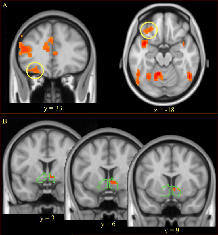
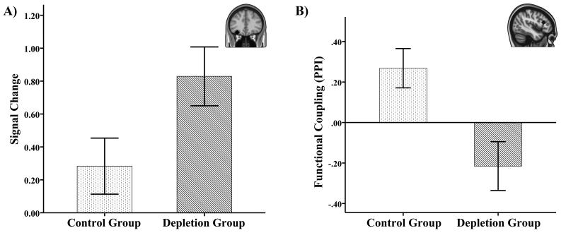
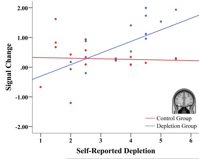
To be successful at self-regulation, individuals must be able to resist impulses and desires. The strength model of self-regulation suggests that when self-regulatory capacity is depleted, self-control deficits result from a failure to engage top-down control mechanisms. Using functional neuroimaging, we examined changes in brain activity in response to viewing desirable foods among 31 chronic dieters, half of whom completed a task known to result in self-regulatory depletion. Compared with nondepleted dieters, depleted dieters exhibited greater food-cue-related activity in the orbitofrontal cortex, a brain area associated with coding the reward value and liking aspects of desirable foods; they also showed decreased functional connectivity between this area and the inferior frontal gyrus, a region commonly implicated in self-control. These findings suggest that self-regulatory depletion provokes self-control failure by reducing connectivity between brain regions that are involved in cognitive control and those that represent rewards, thereby decreasing the capacity to resist temptations.

Keywords: depletion; fMRI; food; orbitofrontal cortex; reward; self-control; self-regulation.


Figures

Figure 1
Figure 1
(A) Brains regions demonstrating greater activity to food versus natural scenes across both depleted and control groups. (p < 0.05, corrected). The yellow circle on the coronal and axial planes indicates the left orbitofrontal cortex. (B) Food-cue related activity in a region of the ventral striatum/nucleus accumbens (p < 0.05 corrected for the volume of the nucleus accumbens). The green outline serves to demarcate the borders of the anatomical volume of the nucleus accumbens that was used for small volume correction.
Figure 2
Figure 2
(A) ROI analysis of the left orbitofrontal cortex (MNI Coordinates: −30,33,−18) demonstrated greater food-cue related activity in depleted as compared to control groups. The orbitofrontal cortex ROI was defined in an unbiased manner from the comparison of food vs. other scenes across both depletion and control groups. (B) Psychophysiolopgical interactions analysis demonstrated reduced context-dependent (food vs. control scenes) functional connectivity between the IFG and OFC in depleted compared to control participants. Separate analysis of each group demonstrated a significant positive context-dependent coupling in the control group (t(14)=2.77, p=0.015) but no difference between food and control scenes in the depleted group (t(15)=1.79, p=0.094). Inset demonstrates the location of the orbitofrontal cortex (a) and inferior frontal gyrus (b) ROI. Error bars indicate SEM.
Figure 3
Figure 3
Individual differences in self-reported depletion following the depletion task predicted subsequent food-cue related activity in the left orbitofrontal cortex in the depleted group (r = 0.591, p = 0.016) but not in the control group (r = − 0.04, p = 0.89). A moderated regression analysis revealed that group was a marginally significant moderator of the relationship between self-reported depletion and OFC activity (β Group x Self-Reported Depletion = 0.331, p = 0.051).

References

    1. Aron AR, Robbins TW, Poldrack RA. Inhibition and the right inferior frontal cortex. Trends in Cognitive Sciences. 2004;8(4):170–177. - PubMed
    1. Baumeister RF, Heatherton T. Self-Regulation Failure: An Overview. Psychological Inquiry. 1996;7(1):1–15.
    1. Beaver JD, Lawrence AD, Van Ditzhuijzen J, Davis MH, Woods A, Calder AJ. Individual differences in reward drive predict neural responses to images of food. The Journal of Neuroscience: The Official Journal of the Society for Neuroscience. 2006;26(19):5160–5166. - PMC - PubMed
    1. Beedie CJ, Lane AM. The Role of Glucose in Self-Control: Another Look at the Evidence and an Alternative Conceptualization. Personality and Social Psychology Review: An Official Journal of the Society for Personality and Social Psychology, Inc. 2011 - PubMed
    1. Berkman ET, Falk EB, Lieberman MD. In the Trenches of Real-World Self-Control. Psychological Science. 2011;22(4):498–506. - PMC - PubMed

Publication types

LinkOut - more resources