. 2014 May;21(5):562-72.
doi: 10.1177/1933719113503406. Epub 2013 Sep 11.

TREM-1 expression is increased in human placentas from severe early-onset preeclamptic pregnancies where it may be involved in syncytialization

Affiliations

TREM-1 expression is increased in human placentas from severe early-onset preeclamptic pregnancies where it may be involved in syncytialization

Ratana Lim et al. Reprod Sci. 2014 May.

Abstract

Preeclampsia, a major cause of maternal and perinatal morbidity and mortality, is thought to be attributable to dysregulation of trophoblast invasion and differentiation. Microarray studies have shown that triggering receptor expressed on myeloid cells (TREM) 1, a cell surface molecule involved in the inflammatory response, is increased in preeclamptic placentas. The aim of this study was to determine the level of TREM-1 expression in severe early-onset preeclamptic placentas and its functional role in trophoblast differentiation. Placenta was obtained from women with severe early-onset preeclampsia (n = 19) and gestationally matched preterm controls placentas (n = 8). The TREM-1 expression was determined by quantitative reverse transcriptase polymerase chain reaction and Western blotting. The effect of TREM-1 small interfering RNA on cell fusion and differentiation was assessed in BeWo cells. The effect of oxygen tension on TREM-1 levels, in basal or forskolin-treated BeWo cells, was also assessed. The TREM-1 was localized to the syncytiotrophoblast layer, and TREM-1 messenger RNA and protein expression was significantly increased in preeclamptic placentas. The BeWo cells treated with forskolin were associated with increased TREM-1 expression. The TREM-1 knockdown inhibited forskolin-induced expression of the differentiation marker β-human chorionic gonadotropin but had no effect on the cell-fusion marker E-cadherin. The increase in TREM-1 expression in BeWo cells treated with forskolin during normoxic conditions was reduced in forskolin-treated cells under hypoxic conditions. In conclusion, TREM-1 is increased in preeclamptic placentas and by forskolin treatment. Knockdown of TREM-1 by RNA interference inhibits cell differentiation but has no effect on cell-cell fusion. Finally, we show that TREM-1 upregulation is attenuated under hypoxic conditions in which cell differentiation is impaired.

Keywords: TREM-1; function; placenta; preeclampsia.


Conflict of interest statement

Declaration of Conflicting Interests: The author(s) declared no potential conflicts of interest with respect to the research, authorship, and/or publication of this article.

Figures

Figure 1.
Figure 1.
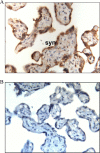
Localization of TREM-1 in human placenta. A, Immunohistochemical localization of TREM-1 in placenta. The TREM-1 staining was in the syncytiotrophoblast (syn) layer. There was no TREM-1 staining in the villous stroma. B, No specific staining for TREM-1 is seen in the negative control for placenta. Magnification 250×. TREM-1 indicates triggering receptor expressed on myeloid cells 1.
Figure 2.
Figure 2.
Increased TREM-1 expression in preeclamptic placentas. The TREM-1 expression in preeclamptic (n = 19) compared to preterm control (n = 8) placentas. A, Gene expression was analyzed by qRT-PCR. The TREM-1 mRNA expression is displayed as mean ± SEM. *P < .05 versus preterm control. B, Representative Western blots and quantitation for TREM-1. Expression levels were confirmed by densitometry. Data are displayed as the mean ± SEM. *P < .05 versus preterm control. mRNA indicates messenger RNA; SEM, standard error of the mean; TREM-1, triggering receptor expressed on myeloid cells 1; qRT-PCR, quantitative reverse transcriptase polymerase chain reaction.
Figure 3.
Figure 3.
Syncytialization of BeWo cells enhances TREM-1 expression. The BeWo cells were incubated in the absence (DMSO control) or presence of 20 μmol/L forskolin for 48 hours (n = 6 independent experiments). A-D, Syncytialization of BeWo was confirmed by decreased E-cadherin and increased β-hCG expression and secretion. A, Gene expression of E-cadherin was analyzed by qRT-PCR, and gene expression is displayed as mean ± SEM. *P < .05 versus control cells. B, Representative Western blot of E-cadherin in cellular lysates in basal and forskolin-treated cells. C, Gene expression of β-hCG was analyzed by qRT-PCR, and gene expression is displayed as mean ± SEM. *P < .05 versus control cells. D, The protein level of β-hCG secreted in media. Data are expression displayed as mean ± SEM. *P < .05 versus control cells. E and F, Syncytialization of BeWo cells enhances TREM-1 expression. E, The TREM-1 gene expression was analyzed by qRT-PCR, and mRNA expression is displayed as mean ± SEM. *P < .05 versus basal cells. F, Representative Western blot of TREM-1 in cellular lysates in basal and forskolin-treated cells. DMSO indicates dimethyl sulfoxide; hCG, gonadotropin hormone; mRNA, messenger RNA; SEM, standard error of the mean; TREM-1, triggering receptor expressed on myeloid cells 1; qRT-PCR, quantitative reverse transcriptase polymerase chain reaction.
Figure 4.
Figure 4.
Loss of TREM-1 is associated with decreased BeWo cell differentiation. The BeWo cells were transfected with 150 nmol/L NS siRNA or TREM-1 siRNA. After 24 hours, cells were incubated in the absence or presence of 20 μmol/L forskolin for an additional 48 hours (n = 6 independent experiments). A and B, Efficiency of siRNA transfection of TREM-1. A, TREM-1 gene expression was analyzed by qRT-PCR, and mRNA expression is displayed as mean ± SEM. *P < .05 versus forskolin-treated NS siRNA-transfected cells. B, Representative Western blots and quantitation for TREM-1 in cellular lysates in forskolin-treated NS and TREM-1 siRNA-transfected cells. C and D, Loss of TREM-1 does not affect E-cadherin expression in syncytialized BeWo cells. C, Gene expression of E-cadherin was analyzed by qRT-PCR, and gene expression is displayed as mean ± SEM. *P < .05 versus forskolin-treated NS siRNA-transfected cells. D, Representative Western blot of E-cadherin in cellular lysates in forskolin-treated NS and TREM-1 siRNA-transfected cells. E and F, Loss of TREM-1 decreases hCG levels in syncytialized BeWo cells. E, Gene expression of β-hCG was analyzed by qRT-PCR, and gene expression is displayed as mean ± SEM. *P < .05 versus forskolin-treated NS siRNA-transfected cells. F, The protein level of β-hCG secreted in media. Data are expression displayed as mean ± SEM. *P < .05 versus forskolin-treated NS siRNA-transfected cells. hCG indicates gonadotropin hormone; mRNA, messenger RNA; NS, nonspecific; SEM, standard error of the mean; siRNA, small interfering RNA; TREM-1, triggering receptor expressed on myeloid cells 1; qRT-PCR, quantitative reverse transcriptase polymerase chain reaction.
Figure 5.
Figure 5.
Hypoxia decreases TREM-1 expression in syncytiotrophoblasts. The BeWo cells were incubated in the absence (DMSO control) or presence of 20 μmol/L forskolin for 48 hours followed by incubation at either 1% or 21% O2 for 24 hours (n = 6 independent experiments). A, The TREM-1 gene expression was analyzed by qRT-PCR, and mRNA expression is displayed as mean ± SEM. *P < .05 versus 21% O2 basal cells. # P < .05 versus 21% O2 forskolin-treated cells. B, Representative Western blot for TREM-1. C, Gene expression of β-hCG was analyzed by qRT-PCR, and gene expression is displayed as mean ± SEM. *P < .05 versus 21% O2 basal cells. # P < .05 versus 21% O2 forskolin-treated cells. D, The protein level of β-hCG secreted in media. Data are expression displayed as mean ± SEM. *P < .05 versus 21% O2 basal cells. # P < .05 versus 21% O2 forskolin-treated cells. DMSO indicates dimethyl sulfoxide; hCG, gonadotropin hormone; mRNA, messenger RNA; SEM, standard error of the mean; TREM-1, triggering receptor expressed on myeloid cells 1; qRT-PCR, quantitative reverse transcriptase polymerase chain reaction.

References

    1. Young BC, Levine RJ, Karumanchi SA. Pathogenesis of preeclampsia. Annu Rev Pathol. 2010;5:173–192. - PubMed
    1. Roberts JM, Hubel CA. The two stage model of preeclampsia: variations on the theme. Placenta. 2009;30(suppl A):S32–S37. - PMC - PubMed
    1. Murphy VE, Smith R, Giles WB, Clifton VL. Endocrine regulation of human fetal growth: the role of the mother, placenta, and fetus. Endocr Rev. 2006;27(2):141–169. - PubMed
    1. Red-Horse K, Zhou Y, Genbacev O, et al. Trophoblast differentiation during embryo implantation and formation of the maternal–fetal interface. J Clin Invest. 2004;114(6):744–754. - PMC - PubMed
    1. Huppertz B, Gauster M. Trophoblast fusion. Adv Exp Med Biol. 2011;713:81–95. - PubMed

Publication types

Substances

LinkOut - more resources