. 2013 Sep 11;3(9):130102.
doi: 10.1098/rsob.130102.

MHF1-2/CENP-S-X performs distinct roles in centromere metabolism and genetic recombination

Affiliations

MHF1-2/CENP-S-X performs distinct roles in centromere metabolism and genetic recombination

Sonali Bhattacharjee et al. Open Biol. .

Retraction in

Abstract

The histone-fold proteins Mhf1/CENP-S and Mhf2/CENP-X perform two important functions in vertebrate cells. First, they are components of the constitutive centromere-associated network, aiding kinetochore assembly and function. Second, they work with the FANCM DNA translocase to promote DNA repair. However, it has been unclear whether there is crosstalk between these roles. We show that Mhf1 and Mhf2 in fission yeast, as in vertebrates, serve a dual function, aiding DNA repair/recombination and localizing to centromeres to promote chromosome segregation. Importantly, these functions are distinct, with the former being dependent on their interaction with the FANCM orthologue Fml1 and the latter not. Together with Fml1, they play a second role in aiding chromosome segregation by processing sister chromatid junctions. However, a failure of this activity does not manifest dramatically increased levels of chromosome missegregation due to the Mus81-Eme1 endonuclease, which acts as a failsafe to resolve DNA junctions before the end of mitosis.

Keywords: DNA helicase; DNA replication; anaphase bridge; centromere; homologous recombination.


Figures
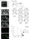
Figure 1.
Figure 1.
MHF functions at the centromere and promotes viability and chromosome segregation independently of Fml1. (a) Spot assay showing the relative growth of strains MCW1221, MCW2080, MCW4639 and MCW4777 on YES agar after 3 days at 30°C. (b) Example cells from a culture of MCW5846 showing the co-localization of Mhf1-GFP with Mis6-mCherry. (c) Schematic of the meiotic chromosome segregation assay. (d) Meiotic chromosome segregation in asci from wild-type (AY167-1D × FO652), fml1Δ (MCW4172 × MCW6196) and mhf2Δ (MCW4777 × MCW5113) homozygous crosses.
Figure 2.
Figure 2.
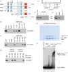
MHF interacts with Fml1. (a) Schematic of Fml1 and the various truncated forms of it used in (b) and (c). The position of the MBP fusion, DNA helicase motifs (blue bars), and region encompassing the site of MHF interaction (red box) are shown. The numbers refer to amino acid positions. (b–d) Western blots showing the amount of His-tagged Mhf2 retained on amylose resin that has been pre-incubated with the indicated MBP-Fml1 fragment (the numbers refer to amino acid positions). (e) Amino acids 670–690 in Fml1 with the mutations tested in (f) indicated. (f) Western blot showing the retention of His-tagged Mhf2 on amylose resin pre-incubated with MBP-Fml1650–690 or its mutant derivatives 1–3. (g) Western blot showing that full-length Fml1AAA fused to MBP fails to retain HisMhf2 on amylose resin. (h) SDS-PAGE analysis of gel filtration fractions 32–38 from the purification of the Fml1576–725–MHF complex. The gel was stained with Coomassie blue. (i) EMSA comparing the ability of MHF and the Fml1576–725–MHF complex to bind linear dsDNA. The amounts of protein are 49 nM (lanes b and f), 98 nM (lanes c and g), 490 nM (lanes d and h) and 980 nM (lanes e and i).
Figure 3.
Figure 3.
MHF functions with Fml1 in the same DNA damage tolerance/repair pathway. (a) Spot assays comparing the growth and genotoxin sensitivities after 5 days of growth at 30°C for (a) strains MCW1221, MCW2080, MCW4639, MCW4777, MCW5127, MCW5790 and MCW5126, and (b) strains MCW1221, MCW2080, MCW5895 and MCW4405.
Figure 4.
Figure 4.
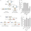
MHF promotes NCO recombination in both mitotic and meiotic cells. (a) Schematic of the plasmid gap repair assay. The filled circle indicates the position of the M26 mutation. (b) Percentage of Ade+ transformants that are COs in strains MCW1193, MCW2096, MCW5345, MCW5346, MCW5790, MCW5983, MCW4893 and MCW6001. Values are means from three experiments ±s.d. (c) Schematic of the meiotic recombination assay showing the different types of CO and NCO recombinants that can be associated with an ade6+ convertant. The filled circles indicate the position of the 3083 and L469 mutations. (d) Percentage of CO associated with GC events at ade6-3083 from wild-type and mutant crosses (see electronic supplementary material, table S1). Values are means from at least six crosses ±s.d. Statistical significance in comparison with wild-type and fml1+::natMX4 is *p < 0.01.
Figure 5.
Figure 5.
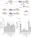
MHF functions with Fml1 to promote RTS1-induced direct repeat recombination. (a) Schematic of the recombination substrate on chromosome III, including the two classes of Ade+ recombinant product. (b) Deletion-type frequencies and (c) conversion-type frequencies in strains MCW4713, MCW3060, MCW4770, MCW5217, MCW5218, MCW5220, MCW4797 and MCW5932. All values are mean±s.d. Statistical significance in comparison with wild-type and fml1+::natMX4 is *p < 0.01 and **p < 0.001.
Figure 6.
Figure 6.
Nuclear localization of Mhf1-GFP in wild-type and fml1 mutant cells. The strains are MCW5846, MCW5963, MCW6152 and MCW6132.
Figure 7.
Figure 7.
Mitotic DNA bridges in wild-type and fml1 mutant cells. (a) Examples of wild-type cells undergoing mitosis. (b) Examples of fml1Δ mutant cells exhibiting a mitotic DNA bridge (top row) and tails (bottom row). (c) Frequency of mitotic DNA bridges/tails and (d) length of mitotic DNA bridges in strains MCW1221, MCW2080, MCW4778 and MCW5895. In all cases, values are mean±s.d. (e) Examples of Mhf1-GFP co-localizing with mitotic DNA bridges/tails in wild-type (MCW5846) and fml1D196N mutant cells (MCW6132).
Figure 8.
Figure 8.
Partial suppression of aberrant chromosome segregation and genotoxin sensitivity in a fml1Δ mus81Δ double mutant by deleting rad51. (a) Examples of aberrant chromosome segregation in septated mus81Δ (MCW1779) and mus81Δ fml1Δ (MCW2428) mutant cells. Arrowheads indicate the division septum. (b) Frequency of aberrant chromosome segregation among septated cells in strains MCW1221, MCW2080, MCW1088, MCW1779, MCW2428 and MCW4490. Values are based on the analysis of approximately 300 septated cells from three independent asynchronously growing cultures. (c) Spot assay comparing the growth and genotoxin sensitivity of strains MCW3792, MCW1235, MCW2428 and MCW4490. Plates were photographed after 5 days growth at 30°C.

References

    1. Heyer WD, Ehmsen KT, Liu J. 2010. Regulation of homologous recombination in eukaryotes. Annu. Rev. Genet. 44, 113–139 (doi:10.1146/annurev-genet-051710-150955) - DOI - PMC - PubMed
    1. Meetei AR, et al. 2005. A human ortholog of archaeal DNA repair protein Hef is defective in Fanconi anemia complementation group M. Nat. Genet. 37, 958–963 (doi:10.1038/ng1626) - DOI - PMC - PubMed
    1. Mosedale G, Niedzwiedz W, Alpi A, Perrina F, Pereira-Leal JB, Johnson M, Langevin F, Pace P, Patel KJ. 2005. The vertebrate Hef ortholog is a component of the Fanconi anemia tumor-suppressor pathway. Nat. Struct. Mol. Biol. 12, 763–771 (doi:10.1038/nsmb981) - DOI - PubMed
    1. Whitby MC. 2010. The FANCM family of DNA helicases/translocases. DNA Repair 9, 224–236 (doi:10.1016/j.dnarep.2009.12.012) - DOI - PubMed
    1. Kee Y, D'Andrea AD. 2010. Expanded roles of the Fanconi anemia pathway in preserving genomic stability. Genes Dev. 24, 1680–1694 (doi:10.1101/gad.1955310) - DOI - PMC - PubMed

Publication types

MeSH terms

LinkOut - more resources