. 2014 Jun;35(6):2779-88.
doi: 10.1002/hbm.22366. Epub 2013 Sep 12.

Intrainsular functional connectivity in human

Affiliations

Intrainsular functional connectivity in human

Talal Almashaikhi et al. Hum Brain Mapp. 2014 Jun.

Abstract

Objectives: The anatomical organization of the insular cortex is characterized by its rich and heterogeneous cytoarchitecture and its wide network of connections. However, only limited knowledge is available regarding the intrainsular connections subserving the complex integrative role of the insular cortex. The aim of this study was to analyze the functional connectivity within- and across-insular subregions, at both gyral and functional levels.

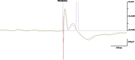
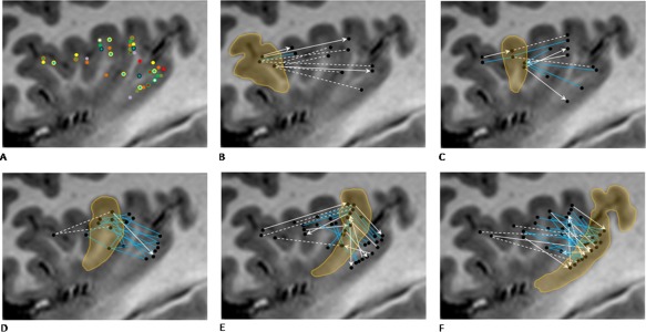
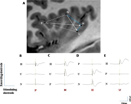
Experimental design: We performed intracerebral electrical stimulation in 10 patients with refractory epilepsy investigated with depth electrodes, 38 of which were inserted in the insula. Bipolar electrical stimulation, consisting of two series of 20 pulses of 1-ms duration, 0.2-Hz frequency, and 1-mA intensity, was delivered at each insular contact. For each stimulated insular anatomical region, we calculated a rate of connectivity, reflecting the proportion of other insular contacts, showing significant evoked potentials.

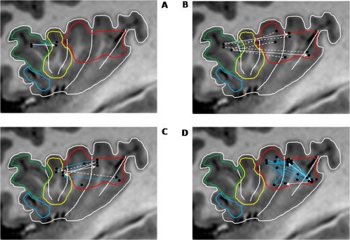
Results: Statistically significant evoked potentials were recorded in 74% of tested connections, with an average latency of 26 ± 3 ms. All insular gyri were interconnected, except the anterior and posterior short gyri. Most connections were reciprocal, showing no clear anterior to posterior directionality. No connection was observed between the right and the left insula.

Conclusions: These findings point to specific features of human insula connectivity as compared to non-Human primates, and remain consistent with the complex integration role devoted to the human insula in many cognitive domains. Periodicals, Inc.

Keywords: evoked potential; functional connectivity; human; insular; intra-cranial electrical stimulation.


Figures

Figure 1
Figure 1
Illustration of EP recorded after insular stimulation. Negative polarity is upward. The green and red color superimposed curves are the average of two 20 trials, showing similar N1 and N2 peaks. The purple curve represents the P‐statistic whose value is 0.001 threshold (i.e., significant response) when reaching the abscissa.
Figure 2
Figure 2
Electrode location and gyral connectivity pattern. A: Electrode location in all 10 patients; different color is used for each patient. B: ASG connectivity. C: MSG connectivity. D: PSG connectivity. E: ALG connectivity. F: PLG connectivity. For graphs B–F, gyrus of interest is encircled and highlighted, blue solid lines indicate bidirectional connectivity, white solid arrows indicate unidirectional connectivity, and dotted white lines indicate lack of detectable connection.
Figure 3
Figure 3
Illustration of the various patterns of connectivity in one patient. A: Connectivity between four insular leads located within the ASG (P), PSG (N), and PLG (H, U). Blue solid lines indicate bidirectional connectivity, white solid arrows indicate unidirectional connectivity, and dotted white lines indicate lack of detectable connection. (BE) EPs evoked by stimulating P, N, H, and U leads, respectively. B: the stimulation of P (ASG) generate EPs only over H (PLG), which are not reciprocal; (C) the stimulation of N (PSG) generate EPs over H and U (PLG), only one of which is reciprocal (H); (D) the stimulation of U (PLG) generate EPs only over H, which are reciprocal; and (E) the stimulation of H (PLG) generate EPs over N (PSG), and U (PLG), both of which are reciprocal.
Figure 4
Figure 4
Connectivity across‐ and within‐main insular functional subregions. A: Connectivity between cognitive and chemical sensory regions. B: Connectivity between cognitive and sensorimotor regions. C: Connectivity between chemical sensory and sensorimotor regions. D: Connectivity within sensorimotor region. Functional regions as delineated by Kurth et al. [2010]: social–emotional (blue), cognitive (green), chemical sensory (yellow), and sensorimotor (red). For graphs (A–D), blue lines indicate bidirectional connectivity, white arrows indicate unidirectional connectivity, and dotted white lines indicate lack of detectable connection.

References

    1. Adolphs R, Tranel D, Damasio AR (2003): Dissociable neural systems for recognizing emotions. Brain Cogn 52:61–69. - PubMed
    1. Aguera PE, Jerbi K, Caclin A, Bertrand O (2011): ELAN: A software package for analysis and visualization of MEG, EEG, and LFP signals. Comput Intell Neurosci 2011:158970. - PMC - PubMed
    1. Alkire MT, White NS, Hsieh R, Haier RJ (2004): Dissociable brain activation responses to 5‐Hz electrical pain stimulation: A high‐field functional magnetic resonance imaging study. Anesthesiology 100:939–946. - PubMed
    1. Anderson JS, Druzgal TJ, Froehlich A, DuBray MB, Lange N, Alexander AL, Abildskov T, Nielsen JA, Cariello AN, Cooperrider JR, Bigler ED, Lainhart JE (2010): Decreased interhemispheric functional connectivity in autism. Cereb Cortex 21:1134–1146. - PMC - PubMed
    1. Augustine JR. (1996): Circuitry and functional aspects of the insular lobe in primates including humans. Brain Res Rev 22:229–244. - PubMed

LinkOut - more resources