. 2013 Sep 24;110(39):E3685-94.
doi: 10.1073/pnas.1311244110. Epub 2013 Sep 11.

Functional role of oligomerization for bacterial and plant SWEET sugar transporter family

Affiliations

Functional role of oligomerization for bacterial and plant SWEET sugar transporter family

Yuan Hu Xuan et al. Proc Natl Acad Sci U S A. .

Abstract

Eukaryotic sugar transporters of the MFS and SWEET superfamilies consist of 12 and 7 α-helical transmembrane domains (TMs), respectively. Structural analyses indicate that MFS transporters evolved from a series of tandem duplications of an ancestral 3-TM unit. SWEETs are heptahelical proteins carrying a tandem repeat of 3-TM separated by a single TM. Here, we show that prokaryotes have ancestral SWEET homologs with only 3-TM and that the Bradyrhizobium japonicum SemiSWEET1, like Arabidopsis SWEET11, mediates sucrose transport. Eukaryotic SWEETs most likely evolved by internal duplication of the 3-TM, suggesting that SemiSWEETs form oligomers to create a functional pore. However, it remains elusive whether the 7-TM SWEETs are the functional unit or require oligomerization to form a pore sufficiently large to allow for sucrose passage. Split ubiquitin yeast two-hybrid and split GFP assays indicate that Arabidopsis SWEETs homo- and heterooligomerize. We examined mutant SWEET variants for negative dominance to test if oligomerization is necessary for function. Mutation of the conserved Y57 or G58 in SWEET1 led to loss of activity. Coexpression of the defective mutants with functional A. thaliana SWEET1 inhibited glucose transport, indicating that homooligomerization is necessary for function. Collectively, these data imply that the basic unit of SWEETs, similar to MFS sugar transporters, is a 3-TM unit and that a functional transporter contains at least four such domains. We hypothesize that the functional unit of the SWEET family of transporters possesses a structure resembling the 12-TM MFS structure, however, with a parallel orientation of the 3-TM unit.

Keywords: evolution; transporter structure.


Conflict of interest statement

The authors declare no conflict of interest.

Figures

Fig. 1.
Fig. 1.
A phylogenic (neighbor-joining) tree of 62 PQ-loop proteins from prokaryotes and eukaryotes. The PQ-loop proteins showed here are listed in Table S1. Arabidopsis thaliana (At), Homo sapiens (Hs) and Bradyrhizobium japonicum (Bj). Protein sequences were aligned with ClustalW, and the phylogenic tree was constructed with MEGA5.1.
Fig. 2.
Fig. 2.
A prokaryotic PQ-loop protein functions as a sucrose transporter. (A) The prokaryotic PQ-loop protein, BjSemiSWEET1, and AtSWEET11 from Arabidopsis were coexpressed with the sucrose FRET sensor FLIPsuc90m∆1V (n ≥ 8) in HEK293T cells. A decrease in biosensor FRET was observed upon addition of sucrose to the culture in AtSWEET11- and BjSemiSWEET1-expressing cells. HEK293T cells transfected with the sensor plasmid only served as a negative control. (B) Sucrose efflux assay from S. elongatus PCC 7942 overexpressing AtSWEET11 and BjSemiSWEET during a 12-h incubation in BG11 media with the indicated NaCl concentration; sucrose was measured from culture supernatants to determine rate of sucrose export (n ≥ 18). Assays were repeated at least three times; error bars indicate SD.
Fig. 3.
Fig. 3.
Coexpression of AtSWEET1 N and C halves or BjSemiSWEET1 in a yeast hexose transporter mutant EBY4000 and homooligomerization of BjSemiSWEET1. (A) The yeast colonies were first grown on SC (Ura- and Trp-) with 2% maltose and then streaked on SC (Ura- and Trp-) with glucose; they grew for 4 d. C122–247 aa, half size of AtSWEET1 contained the second MtN3 motif; N1–121 aa, half size of AtSWEET1 contained the first MtN3 motif and TM4; Neg, empty vectors pDRf1 plus p112AINE as a negative control; Pos, yeast hexose transporter HXT5 as a positive control. The yellow columns represent TMs 1–3, whereas the red columns represent TMs 5–7 of AtSWEET1. The blue columns represent the fourth TM of AtSWEET1, and 3-TMs of BjSemiSWEET are the black columns. (B) Split ubiquitin assay for homooligomerization of BjSemiSWEET1. Interactions of BjSemiSWEET-Cub fusion with BjSemiSWEET1-Nub fusion and a WT variant of Nub (NubWT) or mutant variant of Nub (NubG) were tested. Yeast growth assays on an SC medium (-His, -Trp, and –Leu). (C) Split GFP assays for BjSemiSWEET1 homooligomerization. BjSemiSWEET1-nYFP+BjSemiSWEET1-cCFP is shown in Upper, and BjSemiSWEET1-nYFP+cCFP is shown in Lower. Agrobacterium-mediated transient expression of indicated constructs in N. benthamiana leaves. (Left) Reconstitution of YFP-derived fluorescence. (Right) Bright field images. (Scale bar: 20 μm.)
Fig. 4.
Fig. 4.
AtSWEET interaction network and distribution. (A) Split ubiquitin yeast two-hybrid results of AtSWEET homo- and heterooligomerizations were summarized in Cytoscape. (B) The distribution of 8 homomers and 47 heteromers are shown as a matrix. SWEETs listed horizontally and vertically indicate SWEET-Cub and SWEET-Nub fusions, respectively. Black boxes indicate interactions between two SWEETs, and SWEET-Cub fusions show autoactivation marked with gray boxes.
Fig. 5.
Fig. 5.
Split GFP assays for SWEET homooligomerization. Agrobacterium-mediated transient expression of indicated constructs in N. benthamiana leaves. Reconstitution of YFP-derived fluorescence and bright field images are shown in Left and Right, respectively. Chlorophyll autofluorescence is marked with red, and the white arrows indicate attachment of chloroplast to the plasmolyzed plasma membrane. SWEET4, -8, and -11 YFP samples were plasmolyzed in 4% NaCl. (Scale bars: 20 μm.) Reconstitution of YFP proteins from coexpressing (A and B) SWEET1-nYFP+SWEET1-cCFP, (C and D) SWEET4-nYFP+SWEET4-cCFP, (G and H) SWEET8-nYFP+SWEET8-cCFP, and (I and J) SWEET11-nYFP+SWEET11-cCFP but not from coexpressing (E and F) SWEET6-nYFP+SWEET6-cCFP.
Fig. 6.
Fig. 6.
Functional analysis of mutant AtSWEET1 proteins by complementation of yeast hexose transport defective strain EBY4000: (A) 7-TM with duplication of the first and last three TMs and six mutation sites in three different TM are shown, and (B) growth assays of mutant AtSWEET1 proteins expressed in EBY4000 yeast strain were performed on YNB media containing 2% glucose or maltose. Except for SWEET1 mutants carrying P43T and Y179A, other mutations (P23T, Y57A, G58D, and G180D) in SWEET1 lead to loss of glucose transport activity. Empty (pDRf1) vector and AtSWEET1 were used as the negative and positive controls, respectively. Yeast cells were grown at 30 °C for 4 d.
Fig. 7.
Fig. 7.
Localization of WT and mutant AtSWEET1 proteins in yeast. WT and mutant AtSWEET1-GFP fusion proteins were expressed in the EBY4000 yeast strain. After growth on minimal medium with maltose as the sole carbon source, cells were analyzed by confocal microscopy (SP5). (Left) GFP fluorescence and (Right) bright field images from the following constructs are shown: (A and B) empty vector without GFP expressions, (C and D) AtSWEET1-GFP, (E and F) P23T-GFP, (G and H) P43T-GFP, (I and J) Y57A-GFP, (K and L) Y58D-GFP, (M and N) Y179A-GFP, and (O and P) G180D-GFP. (Scale bar: 10 μm.)
Fig. 8.
Fig. 8.
Inhibition of glucose transport by coexpressing WT and mutant AtSWEET1 proteins in yeast EBY4000. p112AINE empty or p112AINE-SWEET1 vector was cotransformed with pDRf1 empty or pDR-mSWEET1 (P23T, Y57A, G58D, and G180D) vector into yeast strain EBY4000. Growth assays of yeast cells coexpressing WT and mutant AtSWEET1 proteins were performed in YNB media containing 2% glucose or maltose. Coexpression of mutant SWEET1 proteins together with WT inhibited SWEET1 glucose transport activity at different levels, and mutations at either Y57 or G58 showed severe effects.
Fig. 9.
Fig. 9.
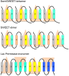
Schematic representation of hypothesized SWEET and SemiSWEET oligomers. Colored boxes indicate TMs, and loops are marked with lines. Numbers in the boxes indicate the order of each TM, and triangles represent functional 3-TM units. Tetramer of SemiSWEET and dimer of SWEET all consist of four 3-TM units, suggesting that 12 helices in consecutive order make functional pores for sugar transport similar as in the 12-TM lactose permease.

References

    1. Walmsley AR, Barrett MP, Bringaud F, Gould GW. Sugar transporters from bacteria, parasites and mammals: Structure-activity relationships. Trends Biochem Sci. 1998;23(12):476–481. - PubMed
    1. Rogers K. Bacteria and Viruses. New York: Britannica Educational Publishing; 2011.
    1. Lalonde S, Wipf D, Frommer WB. Transport mechanisms for organic forms of carbon and nitrogen between source and sink. Annu Rev Plant Biol. 2004;55:341–372. - PubMed
    1. Chen LQ, et al. Sucrose efflux mediated by SWEET proteins as a key step for phloem transport. Science. 2012;335(6065):207–211. - PubMed
    1. Abramson J, et al. Structure and mechanism of the lactose permease of Escherichia coli. Science. 2003;301(5633):610–615. - PubMed

Publication types

MeSH terms