. 2013 Sep 11;33(37):14645-59.
doi: 10.1523/JNEUROSCI.1327-13.2013.

MiR-26b, upregulated in Alzheimer's disease, activates cell cycle entry, tau-phosphorylation, and apoptosis in postmitotic neurons

Affiliations

MiR-26b, upregulated in Alzheimer's disease, activates cell cycle entry, tau-phosphorylation, and apoptosis in postmitotic neurons

Sabrina Absalon et al. J Neurosci. .

Abstract

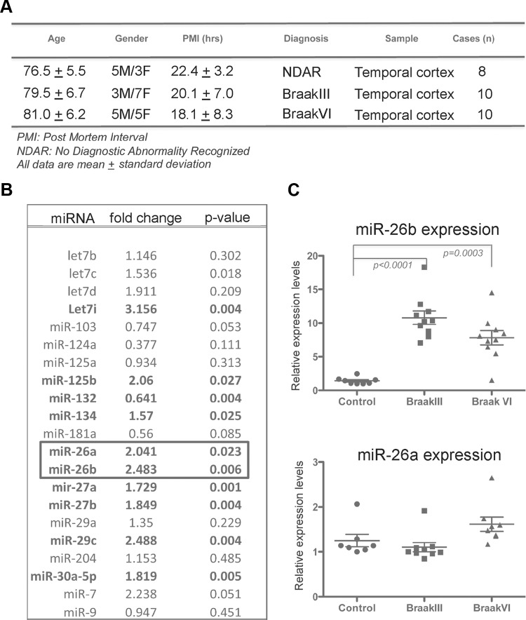
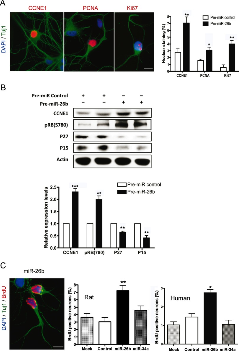
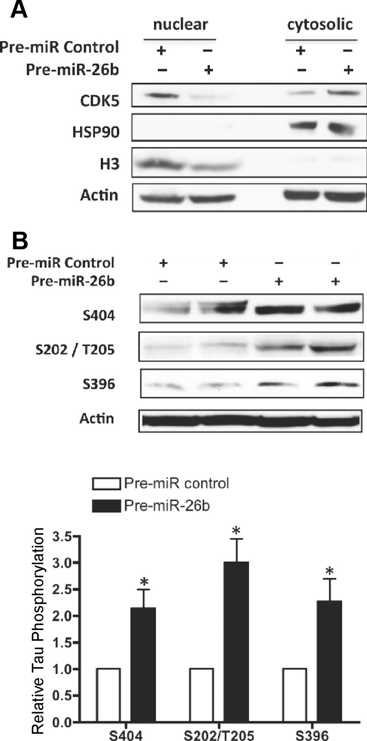
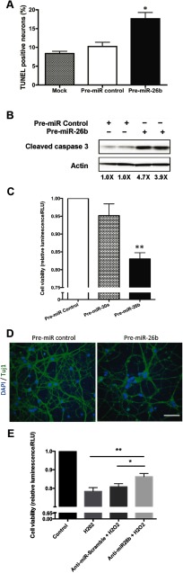
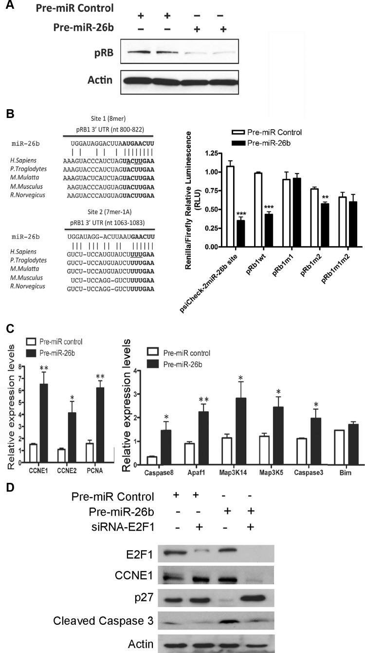
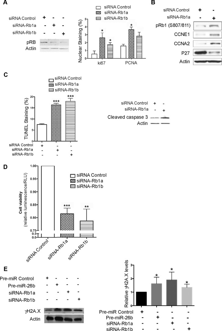
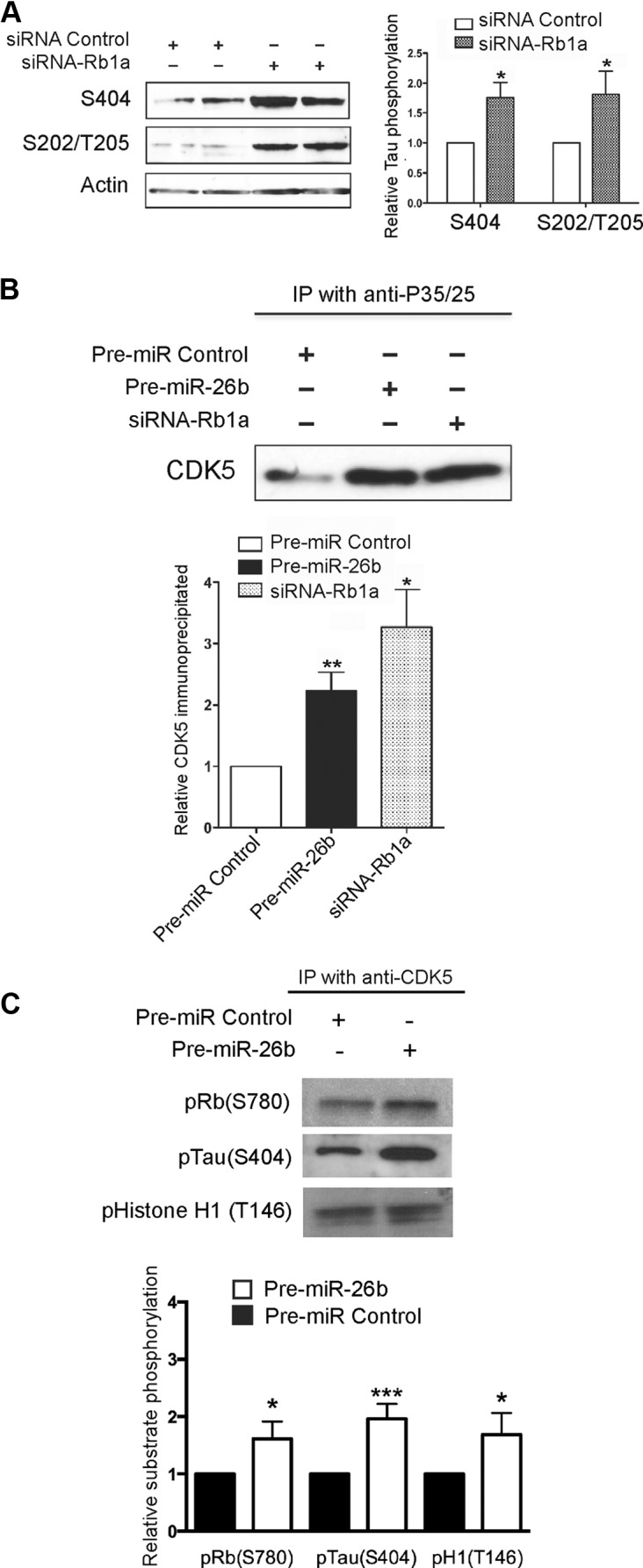
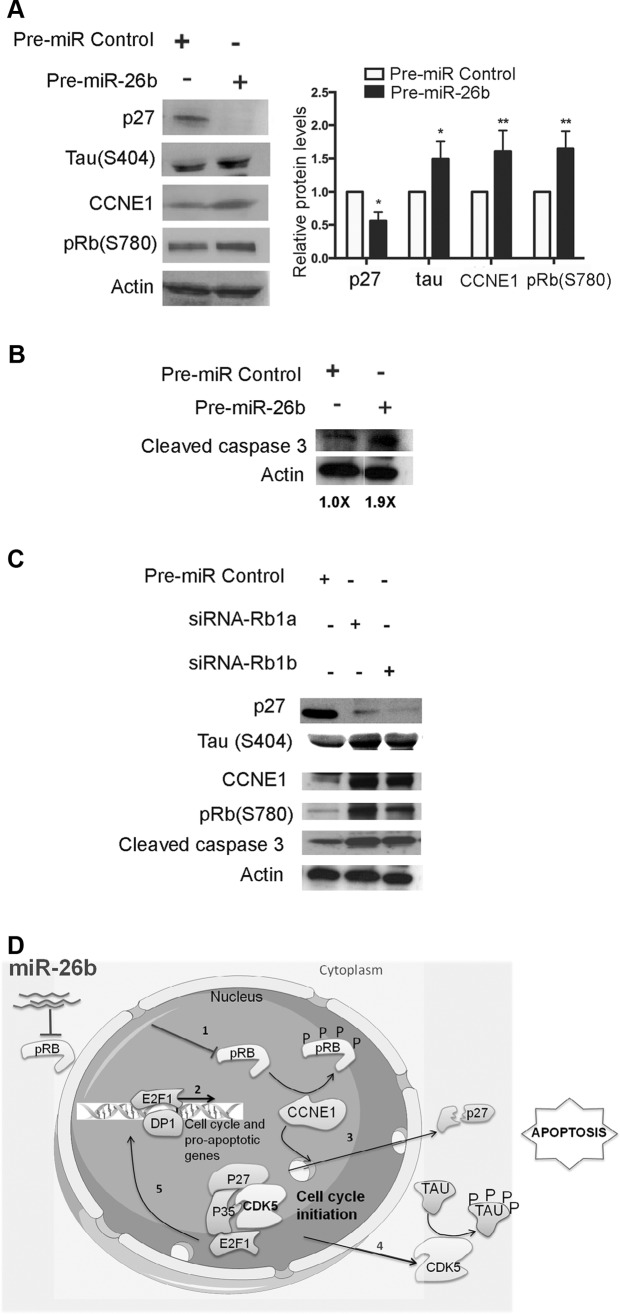
MicroRNA (miRNA) functions in the pathogenesis of major neurodegenerative diseases such as Alzheimer's disease (AD) are only beginning to emerge. We have observed significantly elevated levels of a specific miRNA, miR-26b, in the defined pathological areas of human postmortem brains, starting from early stages of AD (Braak III). Ectopic overexpression of miR-26b in rat primary postmitotic neurons led to the DNA replication and aberrant cell cycle entry (CCE) and, in parallel, increased tau-phosphorylation, which culminated in the apoptotic cell death of neurons. Similar tau hyperphosphorylation and CCE are typical features of neurons in pre-AD brains. Sequence-specific inhibition of miR-26b in culture is neuroprotective against oxidative stress. Retinoblastoma protein (Rb1), a major tumor suppressor, appears as the key direct miR-26b target, which mediates the observed neuronal phenotypes. The downstream signaling involves upregulation of Rb1/E2F cell cycle and pro-apoptotic transcriptional targets, including cyclin E1, and corresponding downregulation of cell cycle inhibitor p27/Kip1. It further leads to nuclear export and activation of Cdk5, a major kinase implicated in tau phosphorylation, regulation of cell cycle, and death in postmitotic neurons. Therefore, upregulation of miR-26b in neurons causes pleiotropic phenotypes that are also observed in AD. Elevated levels of miR-26b may thus contribute to the AD neuronal pathology.


Figures

Figure 1.
Figure 1.
miR-26b expression is upregulated in AD. A, A table summarizes information about the samples used in this study. B, Multiplex miRNA expression analyses were performed on samples from temporal cortex of Braak III (n = 6) and control brains (n = 6). C, Expression of miR-26b (top) and miR-26a (bottom) was tested in control, Braak III, and Braak VI (n = 8–10 per condition) specimens by singleplex qRT-PCR, and relative levels calculated as 2−ΔCt. Uniformly expressed miR-99a was used for normalization. Data represent mean ± SEM with Mann–Whitney test.
Figure 2.
Figure 2.
miR-26b OE induces cell cycle in primary postmitotic neurons. A, miR-26b OE in cortical neurons induces expression of CCNE1, PCNA, and Ki67 compared with neurons transfected with pre-miR control. Typical immunostainings are demonstrated. Histograms depict the number of Tuj1-positive neurons costained with the indicated nuclear markers 5 d post-transfection (Student's t test, two-tailed *p < 0.05 and **p < 0.001; n = 300 neurons/condition). Scale bar, 10 μm. B, Representative Western blot analysis and quantification of four independent experiments demonstrate effects of miR-26b OE on positive (CCNE1 and ppRb1-Ser780) and negative (p27 and p15) regulators of cell cycle (Student's t test, two-tailed **p < 0.001 and ***p < 0.0001, 5 d post-transfection). C, Analysis of BrdU incorporation in neurons by immunostaining for Tuj1 and BrdU demonstrates that OE of miR-26b, but not a control miRNA or miR-34a, induces DNA replication in postmitotic cortical neurons. Representative immunostainings of BrdU+ rat neurons are shown. Histograms depict the percentage of BrdU-positive rat (left) and human (right) neurons (Student's t test, two-tailed *p < 0.05, n = 300). Scale bar, 10 μm.
Figure 3.
Figure 3.
miR-26b OE leads to Cdk5 nuclear export and increased tau-phosphorylation in primary neurons. A, Western blot analysis shows Cdk5 reshuttling from the nuclear to the cytosolic fraction in miR-26b-overexpressing cortical neurons. Histone H3 and Hsp90 were used as markers of nuclear and cytosolic fractions, respectively, and actin as a loading control. B, Western blot analysis shows that miR-26b OE increases tau-phosphorylation at four different epitopes as compared with pre-miR control-transfected neurons. Quantification depicts the densitometric analysis of two experiments (Student's t test, two-tailed *p < 0.05).
Figure 4.
Figure 4.
Primary neurons overexpressing miR-26b show evidence of apoptosis and reduced cell viability. A, Histograms depict the percentage of TUNEL-positive neurons 5 d post-transfection (Student's t test, two-tailed *p < 0.05, n = 3, 300 neurons/condition). B, Western blot analysis and quantification showing that OE of pre-miR-26b leads to an increase in caspase 3 cleavage in postmitotic neurons (Student's t test, two-tailed *p < 0.01, n = 3). C, Cell viability of neurons analyzed by ATP-based assay 5 d post-transfections of pre-miR-26b, pre-miR-30a, or pre-miR control (Student's t test, two-tailed **p < 0.001, n = 3). D, Representative images of neurons transfected with pre-miR-26b or pre-miR control and stained for Tuj1, 7 d post-transfection. Scale bar, 50 μm. E, miR-26b inhibition protects primary neurons against hydrogen peroxide-mediated cytotoxicity. Cell viability of neurons was measured using WST-1 reagent 5 d post-transfection of anti-miR-26b or anti-miR-scramble and 14–16 h post H2O2 treatment (Student's t test, two-tailed **p < 0.005, *p < 0.05, n = 4 with quadruplicate in each experiment).
Figure 5.
Figure 5.
Retinoblastoma is a direct target of miR-26b. A, Western blot analysis shows that miR-26b OE downregulates Rb1 protein in cortical neurons 72 h post-transfection (Student's t test, two-tailed ***p < 0.0001, n = 3). B, Predicted conserved miR-26b binding sites within the Rb1 3′UTR are shown (left). The nucleotides mutated in the Rb1 3′UTR for the luciferase reporter assays are underlined. Relative luciferase reporter activity of cognate miR-26b reporter (left two bars: psiCheck-2 miR-26b site), wild-type (wt), or mutant (m) psiCHECK-2-Rb1 3′UTR constructs cotransfected with a precursor pre-miRNA control or pre-miR-26b (50 nm) in cultures of rat primary neurons (right). OE of miR-26b led to a significant decrease in the relative activity of a wt but not mutated at site 1 construct, as compared with cells transfected with the pre-miR control (Student's t test, two-tailed **p < 0.001 and ***p < 0.0001, n = 3; error bars indicate SEM from 3 independent transfections). C, qRT-PCR analysis of E2F transcriptional targets associated with cell cycle (CCNE1, CCNE2, and PCNA, left) and apoptosis (Caspase 8, Apaf1, Map3K14, Map3K5, Caspase 3, and Bim, right). The quantification reflects expression changes observed 5 d after transfection with pre-miR-26b relative to control. GAPDH served as a normalization housekeeping gene (Student's t test, two-tailed, *p < 0.05, **p < 0.001, n = 3). D, E2F1 downregulation abolishes key effects of miR-26b OE. Western blot analysis of neurons cotransfected with siRNA-E2F1 and either pre-miR-26b or pre-miR control demonstrates a decrease in CCNE1 and cleaved caspase 3 and an increase in p27 in neurons cotransfected with siRNA-E2F1 and pre-miR-26b 5 d post-transfection.
Figure 6.
Figure 6.
Retinoblastoma downregulation in primary cortical neurons by RNAi leads to induction of cell cycle and apoptosis, and mimics the miR-26b OE phenotype. A, Western blot analysis shows that two different siRNAs for Rb1 (siRNA-Rb1a and siRNA-Rb1b) efficiently reduce pRb1 expression 72 h post-transfection (left). Transfections of neuron cultures with these siRNAs lead to increased nuclear staining for both Ki67 and PCNA (right). Histograms depict the percentage of Tuj1+ neurons with the corresponding nuclear staining, counted 5 d post-transfection (Student's t test, two-tailed, *p < 0.05, n = 300 neurons). B, Western blot analysis demonstrates an increase in the expression of cell cycle markers CCNE1 and ppRb1 (S807/811) and a decrease in the expression of the cell cycle inhibitor p27 5 d post-transfection with siRb1. C, Histograms depict the percentage of TUNEL-positive Tuj1+ neurons 5 d post-transfection (Student's t test, two-tailed, ***p < 0.0001, n = 300/3). Western blot analysis (right) shows upregulation of cleaved caspase 3 after transfection with siRb1. D, Cell viability assay demonstrates reduced viability of postmitotic neurons transfected with two siRNAs cognate to Rb1, 5 d post-transfection (Student's t test, two-tailed, **p < 0.001 and ***p < 0.0001, n = 3). E, Western blot analysis (left) of γH2A.X 5 d after transfections of pre-miR-Control, pre-miR-26b, siRNA-Rb1a, or siRNA-Rb1b. Quantification of relative γH2A.X levels shows a significant increase in experimentally transfected neurons as compared with pre-miR control transfected (Student's t test, two-tailed, *p < 0.05, n = 4).
Figure 7.
Figure 7.
Retinoblastoma downregulation by miR-26b or cognate siRNA leads to an increase in tau-phosphorylation and Cdk5 activity in postmitotic neurons. A, Western blot analysis of postmitotic neurons 5 d after siRNA-Rb1 transfection shows an increase of tau-phosphorylation at Ser404 and Ser202/Thr205. Actin serves as a loading control (Student's t test, two-tailed, *p < 0.05, n = 2). B, Immunoprecipitation with anti-p35/25 primary antibody followed by Western blot analysis for Cdk5 reveals an accumulation of Cdk5 associated with p35/p25 in postmitotic neurons transfected with either miR-26b or siRb1 (Student's t test, two-tailed, *p < 0.05 and **p < 0.001, n = 3). C, Immunoprecipitation for Cdk5 followed by in vitro phosphorylation of its substrate peptides Histone H1, Tau, and Rb1, and Western blot analysis with specified phospho-antibodies indicate increased Cdk5 activity in neurons transfected with pre-miR-26b (Student's t test, two-tailed, ***p < 0.0005, *p < 0.01, n = 4).
Figure 8.
Figure 8.
OE of miR-26b or siRNA-mediated knockdown of Rb1 have similar effects in mature cortical neurons transfected at 14 DIV. A, Representative Western blot analysis (left) and quantification of four independent experiments demonstrate that OE of miR-26b increases expression of CCNE1 and phosphorylation of Rb1 at Ser780, and tau at Ser404, whereas p27 is downregulated (two-way ANOVA, *p < 0.05, **p < 0.005, n = 4). B, Western blot analysis showing increased cleavage of caspase 3 5 d post-transfection with pre-miR-26b (Student's t test, two-tailed, *p < 0.05, n = 2). C, Western blot analysis demonstrating that mature cortical neurons transfected with two siRNAs cognate to Rb1 have increased expression of CCNE1 and phosphorylation of Rb1 at Ser780, and tau at Ser404, whereas p27 is downregulated. D, A model of the proposed miR-26b-induced effects on neuronal cell cycle initiation, tau-phosphorylation, Cdk5 activity, and apoptosis.

References

    1. Akashiba H, Matsuki N, Nishiyama N. p27 small interfering RNA induces cell death through elevating cell cycle activity in cultured cortical neurons: a proof-of-concept study. Cell Mol Life Sci. 2006;63:2397–2404. doi: 10.1007/s00018-006-6194-4. - DOI - PMC - PubMed
    1. Andorfer C, Acker CM, Kress Y, Hof PR, Duff K, Davies P. Cell-cycle reentry and cell death in transgenic mice expressing nonmutant human tau isoforms. J Neurosci. 2005;25:5446–5454. doi: 10.1523/JNEUROSCI.4637-04.2005. - DOI - PMC - PubMed
    1. Andrusiak MG, Vandenbosch R, Park DS, Slack RS. The retinoblastoma protein is essential for survival of postmitotic neurons. J Neurosci. 2012;32:14809–14814. doi: 10.1523/JNEUROSCI.1912-12.2012. - DOI - PMC - PubMed
    1. Arendt T, Holzer M, Gärtner U, Brückner MK. Aberrancies in signal transduction and cell cycle related events in Alzheimer's disease. J Neural Transm Suppl. 1998;54:147–158. - PubMed
    1. Bernocco S, Fondelli C, Matteoni S, Magnoni L, Gotta S, Terstappen GC, Raggiaschi R. Sequential detergent fractionation of primary neurons for proteomics studies. Proteomics. 2008;8:930–938. doi: 10.1002/pmic.200700738. - DOI - PubMed

Publication types

MeSH terms

LinkOut - more resources