. 2013 Nov;87(22):12471-80.
doi: 10.1128/JVI.01388-13. Epub 2013 Sep 11.

Antibody recognition of the pandemic H1N1 Influenza virus hemagglutinin receptor binding site

Affiliations

Antibody recognition of the pandemic H1N1 Influenza virus hemagglutinin receptor binding site

Minsun Hong et al. J Virol. 2013 Nov.

Abstract

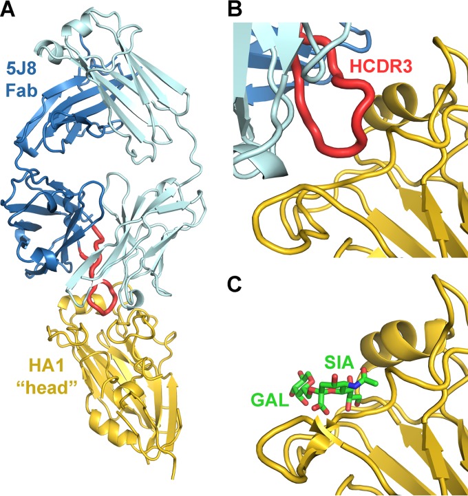
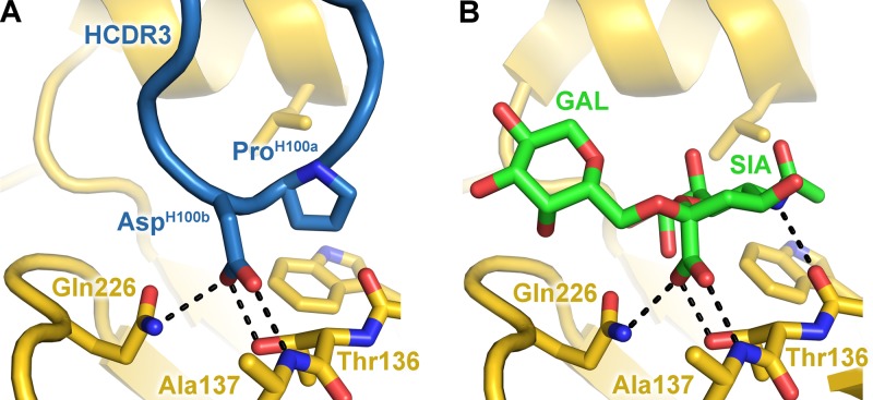
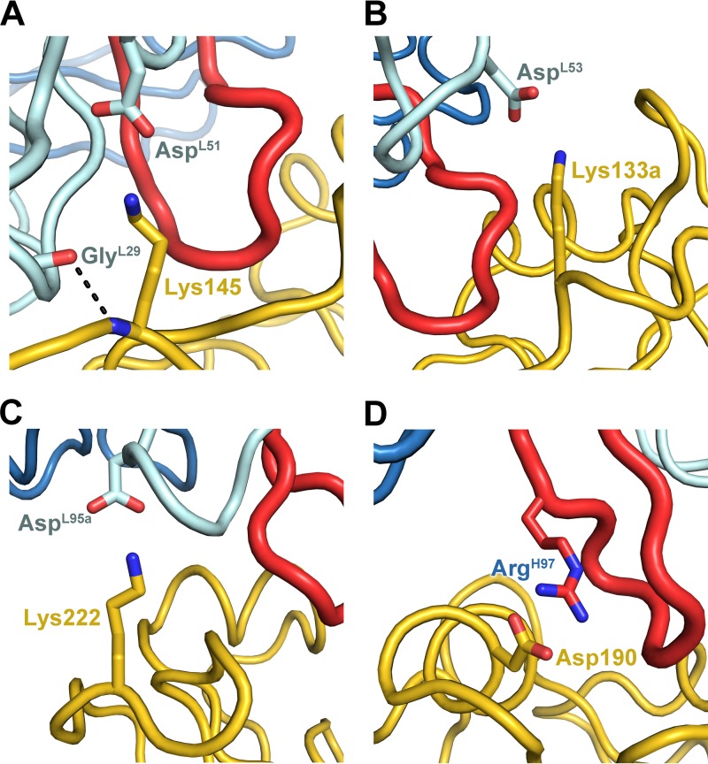
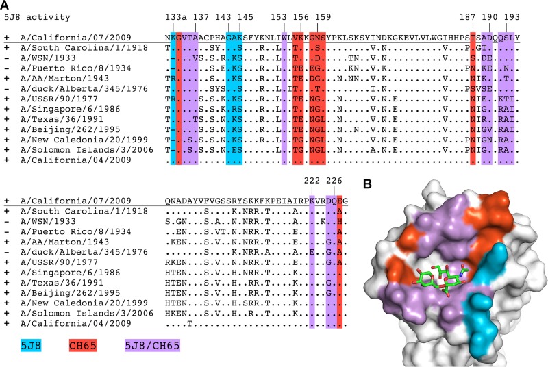
Influenza virus is a global health concern due to its unpredictable pandemic potential. This potential threat was realized in 2009 when an H1N1 virus emerged that resembled the 1918 virus in antigenicity but fortunately was not nearly as deadly. 5J8 is a human antibody that potently neutralizes a broad spectrum of H1N1 viruses, including the 1918 and 2009 pandemic viruses. Here, we present the crystal structure of 5J8 Fab in complex with a bacterially expressed and refolded globular head domain from the hemagglutinin (HA) of the A/California/07/2009 (H1N1) pandemic virus. 5J8 recognizes a conserved epitope in and around the receptor binding site (RBS), and its HCDR3 closely mimics interactions of the sialic acid receptor. Electron microscopy (EM) reconstructions of 5J8 Fab in complex with an HA trimer from a 1986 H1 strain and with an engineered stabilized HA trimer from the 2009 H1 pandemic virus showed a similar mode of binding. As for other characterized RBS-targeted antibodies, 5J8 uses avidity to extend its breadth and affinity against divergent H1 strains. 5J8 selectively interacts with HA insertion residue 133a, which is conserved in pandemic H1 strains and has precluded binding of other RBS-targeted antibodies. Thus, the RBS of divergent HAs is targeted by 5J8 and adds to the growing arsenal of common recognition motifs for design of therapeutics and vaccines. Moreover, consistent with previous studies, the bacterially expressed H1 HA properly refolds, retaining its antigenic structure, and presents a low-cost and rapid alternative for engineering and manufacturing candidate flu vaccines.


Figures

Fig 1
Fig 1
Human monoclonal antibody 5J8 recognizes the HA receptor binding site. (A) Crystal structure of 5J8 Fab in complex with the bacterially expressed Cali07/2009-H1 HA1. The HA1 is colored yellow, the Fab heavy chain is blue, the Fab light chain is light blue, and the HCDR3 is red. 5J8 inserts its HCDR3 into the HA receptor binding site (B) and overlaps with the human α2,6 sialoglycan receptor (PDB accession no. 3UBE) (C).
Fig 2
Fig 2
Degradation of unliganded STF2.HA1 and 5J8 Fab-STF2.HA1 complex. Unliganded STF2.HA1 (∼77 kDa) and 5J8 Fab (∼45 kDa) in complex with STF2.HA1 were incubated at room temperature in the crystallization buffer or the storage buffer (150 mM NaCl, 20 mM Tris [pH 8]). A total of 10 μg of each reaction was quenched at each time point by the addition of nonreducing SDS buffer and was boiled for ∼2 min. Samples were analyzed by SDS-PAGE. Over time, a band at ∼25 kDa appears, which is consistent with the mass of HA1 in the 5J8 Fab-Cali07/2009-H1 complex crystal.
Fig 3
Fig 3
Structural relationships between the bacterially expressed Cali07/2009-H1 HA1 subunit to other HAs. (A) Comparison of the structure of the antigenic sites between the bacterially expressed HA1 (colored yellow) and the full-length baculovirus-expressed Cali04/2009-H1 HA (colored green; PDB accession no. 3LZG) with RMSD values for each site labeled. The Cb antigenic site in our structure is disordered and is not fully modeled. The global-fit RMSD between these HAs is 0.64 Å. A previously solved bacterial HA1 (PDB accession no. 3MLH) is shown in pink and has a global-fit RMSD of 0.6 Å to our structure. (B) Alignment of the HA1 subunits between Cali07/2009-H1 and A/Hong Kong/1/1968 (H3N2) (PDB accession no. 4FP8), colored yellow and gray, respectively. The longer H3 HA1 construct contains additional secondary structural elements of the vestigial esterase domain that extend away from the RBS toward the stem domain as well as two additional disulfide bridges, which are circled and depicted as green sticks. The 5J8 Fab is colored in light and dark blue (heavy and light chains), and its HCDR3 is in red.
Fig 4
Fig 4
Interaction of 5J8 with Cali07/2009-H1 illustrating the neutralizing epitope. (A) Sequence conservation of the 5J8 epitope across human H1 strains. HA residues contacted by 5J8 are represented as yellow sticks. The percent conservation for the most common residue at each position is shown, which is identical to the residues of Cali07/2009-H1. (B) HA is illustrated as a surface in the same orientation as panel A, with the HA contact residues on the surface colored by sequence conservation according to the inserted scale. 5J8 contact residues are labeled and shown as sticks, with the heavy chain in dark blue and the light chain in light blue.
Fig 5
Fig 5
5J8 binds to the HA receptor binding site using receptor mimicry. The carboxylate of AspH100b (A) overlaps with the carboxylate of the α2,6 sialoglycan (PDB accession no. 3UBE) (B) and uses identical hydrogen bonding interactions, which are shown as black dashed lines.
Fig 6
Fig 6
Electrostatic and hydrogen bonding interactions between 5J8 Fab and Cali07/2009-H1 HA1, with hydrogen bonds depicted by dashed lines. The HA is colored yellow, the Fab is colored blue, and the HCDR3 is colored red.
Fig 7
Fig 7
Engineered trimer-stabilized Cali04/2009-H1 HA2 Glu47Gly used for the EM reconstructions. (A) Overview of the HA structure with the HA2 Glu47Gly mutation circled in red. (B) Zoomed-in view of the HA trimer interface. The wild-type HA is colored gray, with Glu47 and the HA1 γ-turn residues (Leu30′ and Glu31′) from the neighboring HA protomer shown as sticks. The trimer-stabilized chains are differentially colored.
Fig 8
Fig 8
EM reconstructions of 5J8 in complex with HA. Negative-stain EM was used to determine volume maps of 5J8 Fab (red) in complex with the full-length HAs (blue) from the A/Singapore/6/1986 (H1N1) and trimer-stabilized A/California/04/2009 (H1N1) strains. The reconstructions show that the HA-antibody interactions described in the 5J8-Cali07/2009-H1 HA1 crystal structure (red and cyan) are recapitulated with the HA trimers. The HA from PDB accession no. 3LZG (blue) was used as a reference structure. Fourier shell correlation (FSC) curves indicate that the resolution for the 5J8-A/Singapore/6/1986 map is 22 Å and for the 5J8-Cali04/2009-H1 map is 23 Å.
Fig 9
Fig 9
Comparison of 5J8 and CH65 contacts on HA. (A) Sequence alignment of the H1 strains tested for binding by biolayer interferometry. (B) Residues contacted by 5J8 (blue), CH65 (red), or both antibodies (violet) are mapped onto the alignment and colored on the surface of Cali07/2009-H1. The sialoglycan (PDB accession no. 3UBE) is represented as sticks. The glyph “.” indicates sequence identity with the Cali07/2009-H1 strain.

References

    1. Wilson IA, Skehel JJ, Wiley DC. 1981. Structure of the haemagglutinin membrane glycoprotein of influenza virus at 3 Å resolution. Nature 289:366–373 - PubMed
    1. Weis W, Brown JH, Cusack S, Paulson JC, Skehel JJ, Wiley DC. 1988. Structure of the influenza virus haemagglutinin complexed with its receptor, sialic acid. Nature 333:426–431 - PubMed
    1. Julien JP, Lee PS, Wilson IA. 2012. Structural insights into key sites of vulnerability on HIV-1 Env and influenza HA. Immunol. Rev. 250:180–198 - PMC - PubMed
    1. Xu R, Ekiert DC, Krause JC, Hai R, Crowe JE, Jr, Wilson IA. 2010. Structural basis of preexisting immunity to the 2009 H1N1 pandemic influenza virus. Science 328:357–360 - PMC - PubMed
    1. Whittle JR, Zhang R, Khurana S, King LR, Manischewitz J, Golding H, Dormitzer PR, Haynes BF, Walter EB, Moody MA, Kepler TB, Liao HX, Harrison SC. 2011. Broadly neutralizing human antibody that recognizes the receptor-binding pocket of influenza virus hemagglutinin. Proc. Natl. Acad. Sci. U. S. A. 108:14216–14221 - PMC - PubMed

Publication types

MeSH terms