. 2013 Dec;87(24):13094-106.
doi: 10.1128/JVI.00704-13. Epub 2013 Sep 11.

Nucleolin interacts with the dengue virus capsid protein and plays a role in formation of infectious virus particles

Affiliations

Nucleolin interacts with the dengue virus capsid protein and plays a role in formation of infectious virus particles

Corey A Balinsky et al. J Virol. 2013 Dec.

Abstract

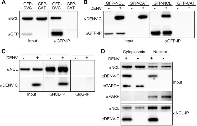
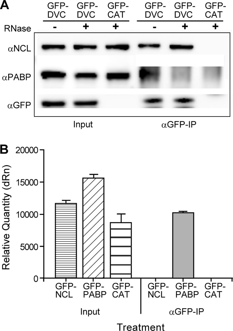
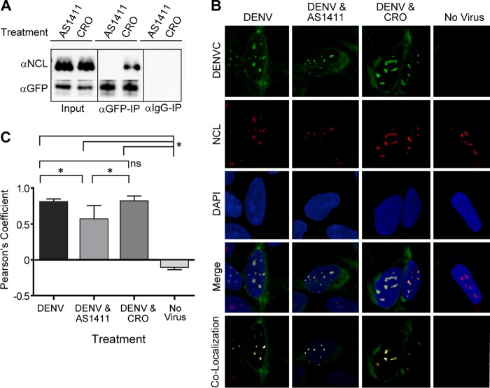
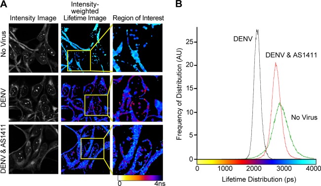
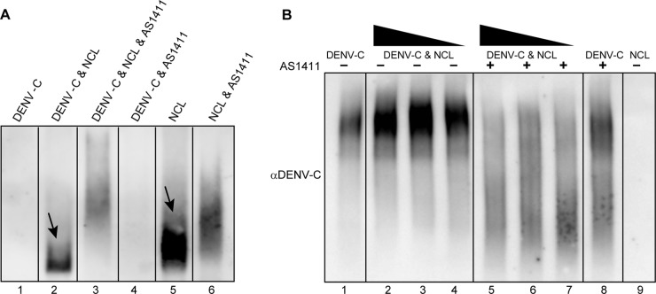
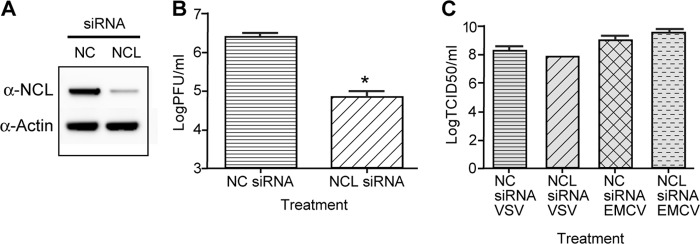
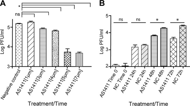
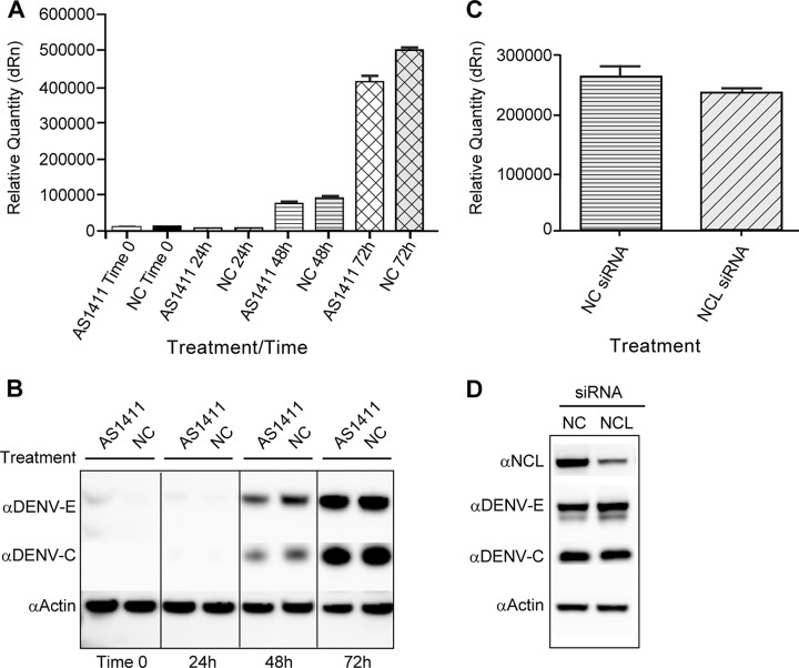
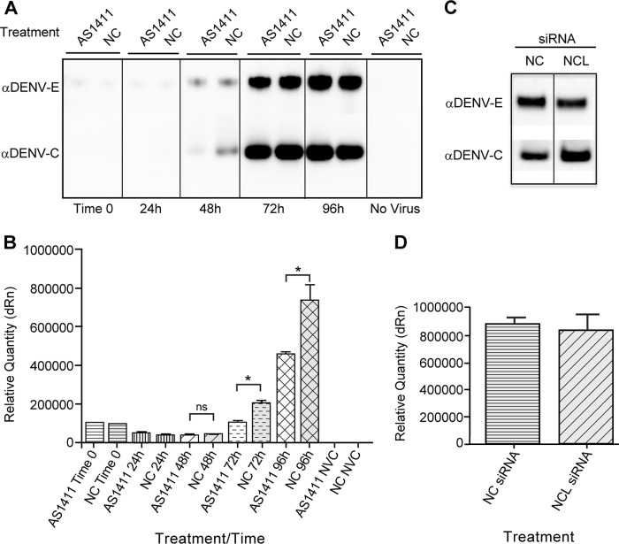
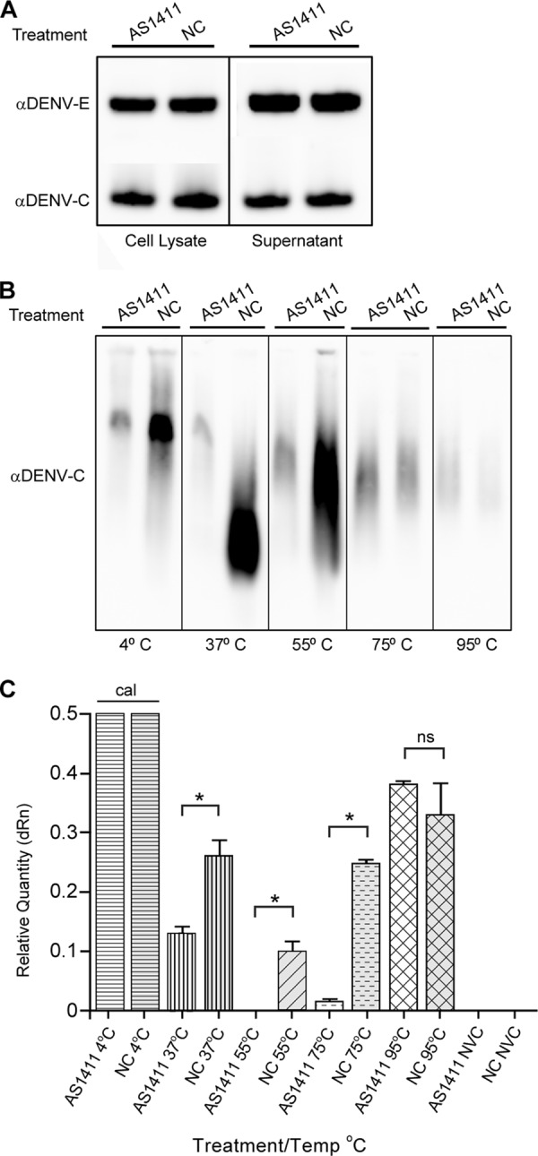
Dengue virus (DENV) is a mosquito-transmitted flavivirus that can cause severe disease in humans and is considered a reemerging pathogen of significant importance to public health. The DENV capsid (C) protein functions as a structural component of the infectious virion; however, it may have additional functions in the virus replicative cycle. Here, we show that the DENV C protein interacts and colocalizes with the multifunctional host protein nucleolin (NCL). Furthermore, we demonstrate that this interaction can be disrupted by the addition of an NCL binding aptamer (AS1411). Knockdown of NCL with small interfering RNA (siRNA) or treatment of cells with AS1411 results in a significant reduction of viral titers after DENV infection. Western blotting and quantitative RT-PCR (qRT-PCR) analysis revealed no differences in viral RNA or protein levels at early time points postinfection, suggesting a role for NCL in viral morphogenesis. We support this hypothesis by showing that treatment with AS1411 alters the migration characteristics of the viral capsid, as visualized by native electrophoresis. Here, we identify a critical interaction between DENV C protein and NCL that represents a potential new target for the development of antiviral therapeutics.


Figures

Fig 1
Fig 1
NCL interacts with DENV C. (A) Co-IP of HEK293 cells transfected with an expression vector containing the DENV C gene (GFP-DVC) or a negative-control vector (GFP-CAT). Co-IP was performed using anti-GFP (αGFP) antibody, and Western blots were stained using antibodies to NCL or GFP. (B) Reciprocal co-IP of HEK293 cells transfected with expression vectors containing NCL (GFP-NCL) or GFP-CAT. Cells were infected with DENV for 48 h or left uninfected. Co-IPs were performed as described for panel A, and Western blots were stained with antibodies to DENV C or GFP. (C) Endogenous co-IP of HEK293 cells infected with DENV for 48 h or left uninfected. Co-IP was performed using αNCL antibody or a nonspecific mouse IgG antibody, and Western blots were stained with antibodies to DENV C or NCL. (D) Endogenous co-IP of HEK293 cells infected with DENV for 48 h or left uninfected. After nuclear/cytoplasmic fractionation, co-IP was performed using αNCL antibody or a nonspecific mouse IgG antibody, and Western blots were stained with antibodies to DENV C or NCL. Antibodies to GAPDH and poly(ADP-ribose) polymerase (PARP) were used as markers for the cytoplasmic and nuclear fractions, respectively. All Western blots are representative of three or more independent experiments.
Fig 2
Fig 2
NCL interaction with DENV C is RNA independent. (A) Co-IP of HEK293 cells transfected with either GFP-DVC or GFP-CAT. Cell lysates were treated with RNase A or left untreated, and co-IP was performed using αGFP antibody as previously described. Western blots were stained using antibodies to NCL, PABP, or GFP. The Western blots are representative of three independent experiments. (B) RNA-IP of HEK293 cells transfected with GFP-NCL, GFP-PABP, or GFP-CAT expression vectors. Co-IP was performed using αGFP antibody, and RNA was extracted from the input or co-IP sample. Samples were analyzed by qRT-PCR using DENV forward and reverse primers. The data are representative of two independent experiments performed in triplicate. dRn, change in the normalized reporter signal.
Fig 3
Fig 3
Treatment with AS1411 blocks interaction between NCL and DENV C and affects colocalization. (A) Co-IP of HEK293 cells transfected with expression vector GFP-DVC. The cells were treated with AS1411 or negative-control CRO (10 μM). Co-IP was performed as previously described, and the Western blots were stained with antibodies to NCL or GFP. (B) Confocal microscopy of HEK293 cells left uninfected or infected with DENV, followed by no treatment or treatment with AS1411 or CRO (10 μM). Samples were examined for localization of DENV C (green) and NCL (red). The nucleus was stained with DAPI (blue). Colocalization of DENV C and NCL is shown in white. (C) Colocalization coefficients of DENV C and NCL in uninfected cells or cells infected with DENV and left untreated or treated with AS1411 or CRO (10 μM), as determined by Pearson's linear correlation coefficient. Significance was determined using a P value of 0.01 (indicated by an asterisk [*]) from 10 images collected from two independent experiments. ns, not significant. The error bars indicate SD.
Fig 4
Fig 4
Treatment with AS1411 affects in vivo interactions between DENV C and NCL as determined by FRET-FLIM analysis. (A) Representative image of FRET-FLIM showing the image intensity, intensity-weighted lifetime, and selected region of interest in uninfected HEK293 cells and cells infected with DENV and left untreated or treated with AS1411 (10 μM). The image is representative of 7 images from each treatment group collected from two independent experiments. The color table corresponds to fluorescence lifetime values. (B) Lifetime histogram of FRET-FLIM of uninfected HEK293 cells, DENV-infected cells, and DENV-infected cells treated with AS1411. The histogram was calculated from 7 images collected from two independent experiments showing lifetime distribution in picoseconds versus frequency of distribution in arbitrary units (AU).
Fig 5
Fig 5
Treatment with AS1411 affects interactions between purified recombinant DENV C and purified NCL. (A) Purified recombinant DENV C protein was incubated for 1 h either alone or in the presence of purified NCL, with or without AS1411 (10 μM). Samples were analyzed by native PAGE, followed by Western blotting of purified NCL protein. The arrows indicate bands corresponding to NCL. (B) Purified recombinant DENV C protein was incubated with a 1,000-fold lower concentration (by molar ratio) of purified NCL or with 10- or 100-fold dilutions thereof, with or without AS1411. Samples were analyzed by native PAGE, followed by Western blotting of recombinant DENV C protein.
Fig 6
Fig 6
siRNA knockdown of NCL results in decreased titers of DENV. HEK293 cells were treated with NCL siRNA or NC siRNA. Twenty-four hours after siRNA treatment, the cells were infected with DENV at an MOI of 0.1, and samples were collected 72 h after infection. (A) Western blots were performed on cell lysates using antibody to NCL to verify siRNA knockdown and an antibody to actin as a loading control. (B) The titer of DENV collected from NC and NCL siRNA groups was determined in duplicate on Vero cells. Three independent experiments were used to determine significance (P < 0.01). (C) HEK293 cells were treated with NC or NCL siRNA as described for panel A. The cells were then infected with either EMCV (MOI, 0.001) or VSV (MOI, 0.001) for 24 h. The titers of samples were determined in quadruplicate on Vero cells using the limiting-dilution method, and the 50% tissue culture infectious dose (TCID50) was calculated. Two independent experiments were used to determine significance (P < 0.01). The error bars indicate SD.
Fig 7
Fig 7
Treatment with AS1411 results in decreased titers of DENV. (A) Virus titers from HEK293 cells infected with DENV (MOI, 0.1) and treated with AS1411 at 1 μM, 3 μM, 4 μM, 5 μM, or 7 μM or with negative-control CRO at 7 μM per ml. Samples were collected at 72 h postinfection, and the titers were determined in duplicate on Vero cells (P < 0.01). (B) Virus titers from HEK293 cells infected with DENV (MOI, 3), followed by treatment with AS1411 or the NC. Samples were collected at time zero and at 24-h intervals for 72 h postinfection. The titers of samples were determined in duplicate on Vero cells. The data are representative of three independent experiments (P < 0.01). The error bars indicate SD.
Fig 8
Fig 8
NCL does not significantly affect DENV RNA replication or protein translation. (A) qRT-PCR of viral RNA extracted from DENV-infected (MOI, 3) HEK293 cells after treatment with AS1411. Samples were analyzed using primers to DENV and normalized to GAPDH. (B) Western blot of cell lysates collected from DENV-infected cells, followed by treatment with AS1411 or the NC and collection at the indicated time points. The Western blots were stained with antibodies to the DENV C and DENV E proteins, as well as with actin. The Western blots are representative of three independent experiments. (C) qRT-PCR of viral RNA extracted from HEK293 cells treated with siRNA to NCL or nonspecific siRNA, followed by infection with DENV (MOI, 3) for 72 h. Samples were analyzed using primers to DENV and normalized to GAPDH. (D) Western blot of HEK293 cells treated with siRNA to NCL or the NC, followed by infection with DENV (MOI, 3). Samples were collected at 72 h postinfection and examined by Western blotting for NCL, DENV C, DENV E, and actin. The error bars indicate SD.
Fig 9
Fig 9
NCL does not significantly affect release of DENV particles. (A) Western blot of HEK293 cells infected with DENV (MOI, 3), followed by treatment with AS1411 or the NC. Cell supernatants were collected at time zero and at 24-h intervals until 96 h postinfection. Virus was purified through a sucrose cushion, and samples were analyzed by SDS-PAGE, followed by Western blotting of DENV C and DENV E proteins. The data are representative of three independent experiments. (B) qRT-PCR of viral RNA extracted from cell supernatants of DENV-infected HEK293 cells (as described for panel A) and no-virus control (NVC). Samples were analyzed using primers to DENV and normalized to norovirus G2 RNA added to the sample prior to extraction. The data are representative of two independent experiments performed in triplicate. (C) Western blot of HEK293 cells treated with siRNA to NCL or the NC, followed by infection with DENV (MOI, 3). Cell supernatants were collected at 72 h postinfection. Virus was purified through a sucrose cushion, and samples were analyzed by SDS-PAGE, followed by Western blotting of DENV C and DENV E proteins. (D) qRT-PCR of viral RNA extracted from cell supernatants of DENV-infected HEK293 cells after treatment with siRNA for NCL or negative-control siRNA. Samples were analyzed using primers to DENV and normalized to norovirus G2 RNA added to the sample prior to extraction. The data are representative of two independent experiments performed in triplicate. The error bars indicate SD.
Fig 10
Fig 10
NCL affects DENV capsid migration characteristics. HEK293 cells were infected with DENV (MOI, 3), followed by treatment with AS1411 or the negative control. Cell supernatants were collected at 72 h postinfection, and virus was purified through a sucrose cushion. (A) Sucrose-purified virus was examined by SDS-PAGE, followed by Western blotting for DENV C and E proteins. (B) The sucrose-purified samples were also incubated for 10 min at the indicated temperatures and run on a native AGE gel. Samples were analyzed by Western blotting for DENV C protein. (C) qRT-PCR of sucrose-purified virus after incubation at the indicated temperatures and NVC. Viral RNA was detected by qRT-PCR using primers to DENV and normalized to the 4°C input samples. The data are representative of two independent experiments performed in triplicate. The error bars indicate SD.

References

    1. WHO 2006. Scientific working group report on dengue. WHO reference number TDR/SWG/08. WHO, Geneva, Switzerland
    1. Guzman MG, Halstead SB, Artsob H, Buchy P, Farrar J, Gubler DJ, Hunsperger E, Kroeger A, Margolis HS, Martínez E, Nathan MB, Pelegrino JL, Simmons C, Yoksan S, Peeling RW. 2010. Dengue: a continuing global threat. Nat. Rev. Microbiol. 8:S7–S16 - PMC - PubMed
    1. Kyle JL, Harris E. 2008. Global spread and persistence of dengue. Annu. Rev. Microbiol. 62:71–92 - PubMed
    1. Perera R, Kuhn RJ. 2008. Structural proteomics of dengue virus. Curr. Opin. Microbiol. 11:369–377 - PMC - PubMed
    1. Rodenhuis-Zybert IA, Wilschut J, Smit JM. 2010. Dengue virus life cycle: viral and host factors modulating infectivity. Cell. Mol. Life Sci. 67:2773–2786 - PMC - PubMed

Publication types

LinkOut - more resources