. 2013 Jun:30:77-93.
doi: 10.3767/003158513X666268. Epub 2013 Mar 13.

A comprehensive molecular phylogeny of the Mortierellales (Mortierellomycotina) based on nuclear ribosomal DNA

Affiliations

A comprehensive molecular phylogeny of the Mortierellales (Mortierellomycotina) based on nuclear ribosomal DNA

L Wagner et al. Persoonia. 2013 Jun.

Abstract

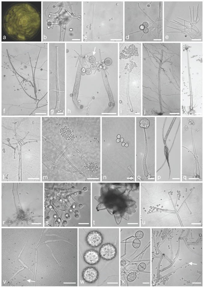
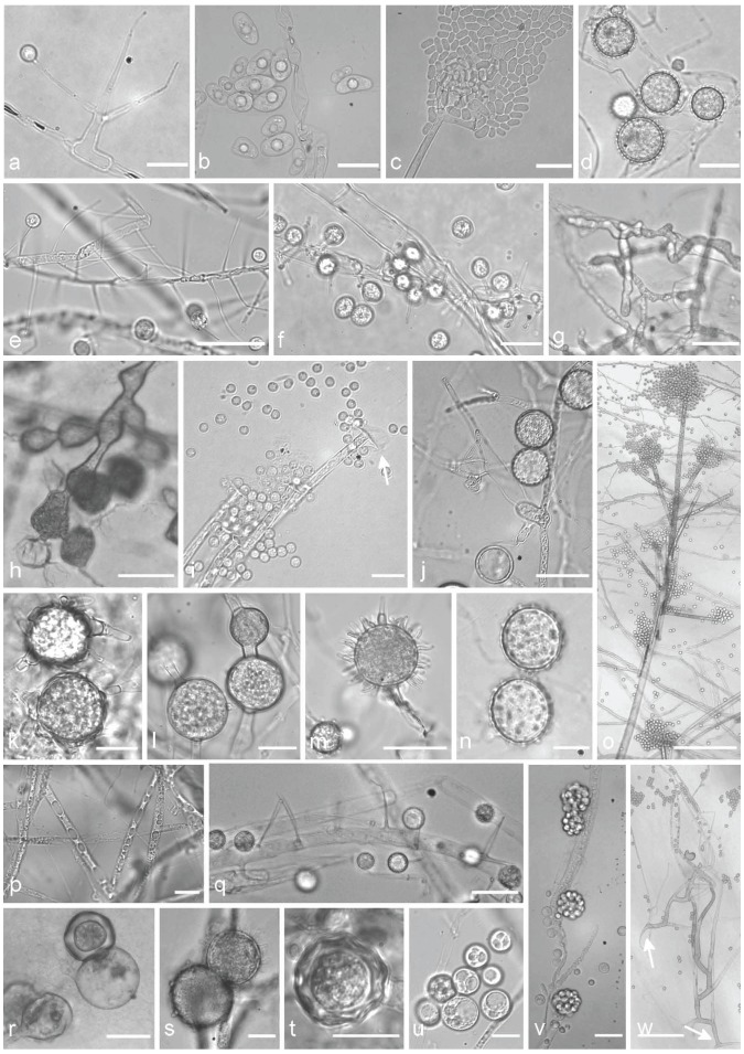
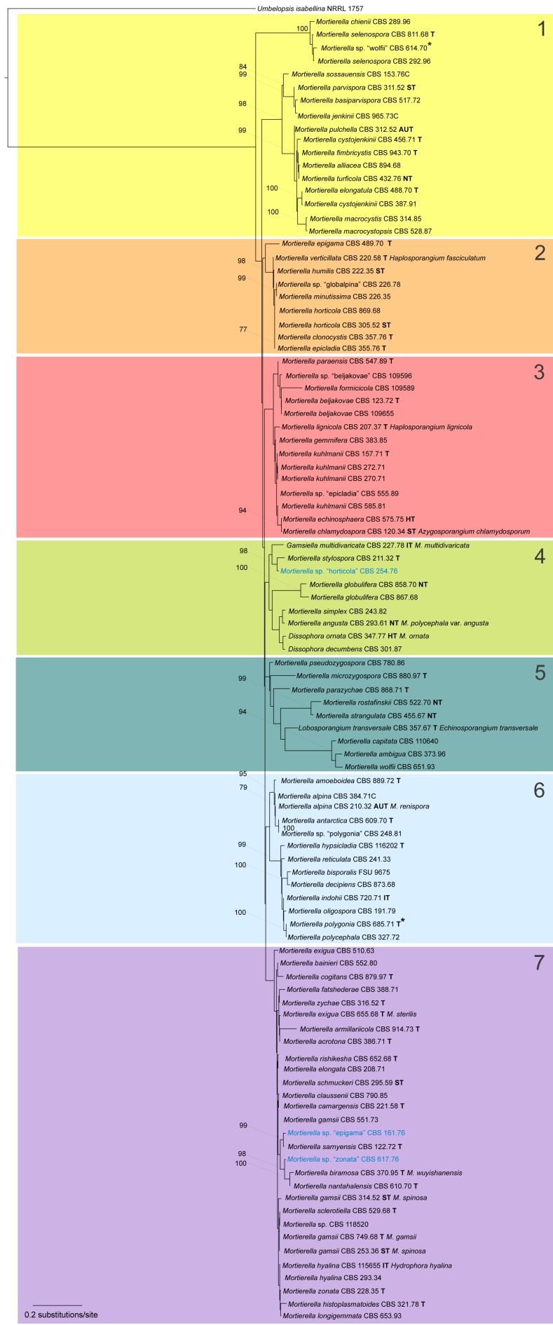
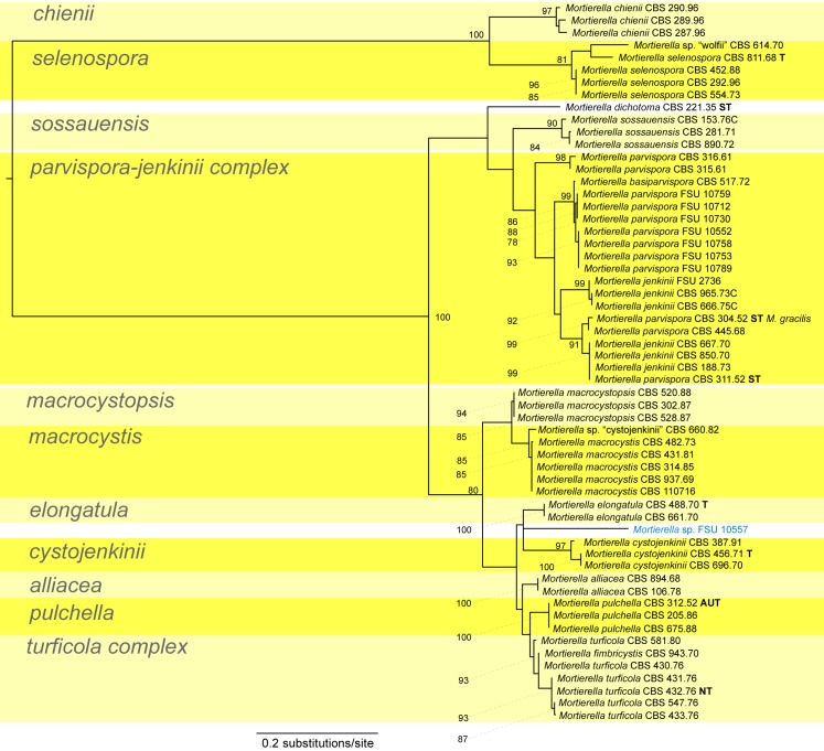
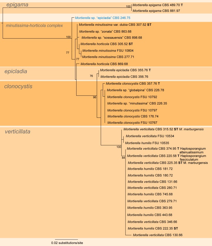
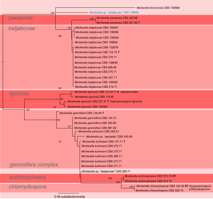
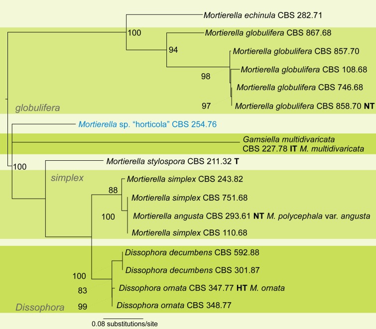
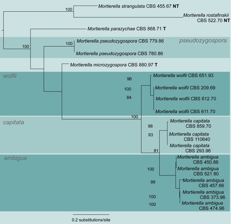
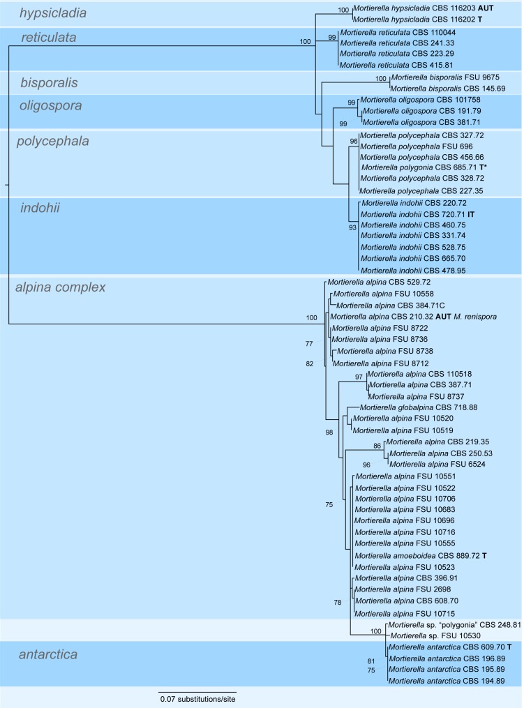
The basal fungal order Mortierellales constitutes one of the largest orders in the basal lineages. This group consists of one family and six genera. Most species are saprobic soil inhabiting fungi with the ability of diverse biotransformations or the accumulation of unsaturated fatty acids, making them attractive for biotechnological applications. Only few studies exist aiming at the revelation of the evolutionary relationships of this interesting fungal group. This study includes the largest dataset of LSU and ITS sequences for more than 400 specimens containing 63 type or reference strains. Based on a LSU phylogram, fungal groups were defined and evaluated using ITS sequences and morphological features. Traditional morphology-based classification schemes were rejected, because the morphology of the Mortierellales seems to depend on culture conditions, a fact, which makes the identification of synapomorphic characters tedious. This study belongs to the most comprehensive molecular phylogenetic analyses for the Mortierellales up to date and reveals unresolved species and species complexes.

Keywords: Zygomycetes; Zygomycota; internal transcribed spacer; large subunit ribosomal DNA; taxonomic revision.


Figures

Fig. 1.
Fig. 1.
Typical morphological structures of different isolates of the Mortierellales, which are suitable for species delimitation. a. M. zychae CBS 316.52, macroscopic shape of a growing culture with the typical zonate growth; b. M. hypsicladia CBS 116202, acrotonous branching of a sporangiophore; c. M. epicladia CBS 355.76, sporangiophore and sporangiospores; d. M. zonata CBS 228.35, basitonous branched sporangiophore with sporangioles; e. Gamsiella multidivaricata CBS 227.78, typical branched sporangiophores; f. M. elongata FSU 9721, basitonous branched sporangiophore; g. M. alpina FSU 2698, sporangiophore; h. M. polycephala FSU 867, sporangiospores with sporangia (arrow) and sporangiospores; i. Mortierella cf. wolfii CBS 614.70, sporangiophore with elongated sporangiospores; j. M. parvispora FSU 10759, sporangiophores; k. M. hypsicladia CBS 116202, typical sporangiophore with rhizoid; l. Mortierella cf. wolfii CBS 614.70, acrotonous branching of a sporangiophore; m. Mortierella sp. FSU 10557, sporangiophore and sporangiospores; n. M. paraensis CBS 547.89, tips of a sporangiophore with a pseudocolumella and sporangiospores; o. M. alpina FSU 2698, sporangiophore with unmatured sporangia; p. M. nanthalensis CBS 610.70, typical rhizoid of a sporangiophore; q. M. wolfii CBS 651.93, sporangiospores with unusual remain of the sporangia cover (arrow); r. M. strangulata CBS 455.67, rhizoid of the sporangiophore; s. Gamsiella multidivaricata CBS 227.78, sporangiophores with sporangioles; t. Lobosporangium transversale CBS 357.67, typical sporangia, arranged in clusters, containing numerous spherical sporangiospores; u. M. gamsii FSU 10538, acrotonous branching of a sporangiophore and sporangiospores; v. Dissophora decumbens CBS 592.88, septate sporangiophores along a hypha and sporangiospore (arrow); w. M. polycephala FSU 867, stylospores; x. Gamsiella multidivaricata CBS 227.78, sporangiola containing spores; y. M. kuhlmanii CBS 157.71, branching pattern of the basitonous part of the sporangiophore and elongated sporangiospores, pseudocolumella. — Scale bars: b, c, s–u, x = 30 μm; d, e, i = 20 μm; f, j, k, p = 100 μm; g, n, o, w = 10 μm; h, l, m, q, r, v, y = 50 μm.
Fig. 2.
Fig. 2.
Typical morphological structures of different isolates of the Mortierellales, which are suitable for species delimitation. a. M. verticillata CBS 315.52, sporangiophore with a sporangiola; b. M. elongata FSU 9721, elongated sporangiospores containing central oil droplets; c. M. wolfii CBS 651.93, cracked sporangia releasing sporangiospores, on acrotonous branched tip of the sporangiophore; d. M. indohii CBS 720.71, stylospores; e. M. schmuckeri CBS 295.59, sporangiophores alongside a hypha with sporangiola; f. M. claussenii CBS 294.59, sporangiophores along a hypha with sporangiola; g. M. clonocystis CBS 357.76, typical swollen hyphae; h. M. zychae FSU 719, typical swollen hyphae arranged in clusters; i. M. parvispora FSU 10759, tip of a sporangiophore, sporangia leaving a collar (arrow), globose sporangiospores; j. M. lignicola CBS 207.37, sporangiophores, sporangiola (arrow 1), stylospores (arrow 2); k. M. exigua CBS 655.68, chlamydospores with typical outgrowing hyphae; l. M. gemmifera CBS 134.45, chlamydospores; m. M. hypsicladia CBS 116202, stylospores with projections; n. M. polygonia CBS 685.71, stylospores; o. M. nanthalensis CBS 610.70, acrotonous branching part of a sporangiophore; p. M. alpina FSU 2698, oil droplets containing hypha; q. M. camargensis CBS 221.58, sporangiophores along a hypha with sporangiola; r. M. epigama CBS 489.70, zygospores; s. M. echinosphaera CBS 575.75, chlamydospores; t. M. microszygospora CBS 880.97, microzygospore; u. M. camargensis CBS 221.58, oil droplets containing spheric sporangiola; v. Dissophora decumbens CBS 592.88, sporangiophores with sporangia; w. M. paraensis CBS 547.89, two sporangiophores with typical basitonous branchings (arrows mark the basal part). — Scale bars: a, b, i, n, p, r, u = 10 μm; c, j, q = 20 μm; d, e, g, h, m, v = 30 μm; f, k, l, s, t = 15 μm; o = 250 μm; w = 100 μm.
Fig. 3.
Fig. 3.
Maximum Likelihood analysis based on 781 aligned nucleotides of the D1/D2 domain of the large subunit (LSU, 28S) rDNA from 101 taxa (100 ingroup taxa of the Mortierellales and 1 outgroup taxon Umbelopsis as member of the Mucorales, Meyer & Gams 2003). The phylogram based on a MAFFT-Alignment (L-ins-I). Node supports above 75 % is given. The tree defines 7 groups: groups 1–7, which are more profoundly analysed in individual analyses based on the ITS1-5.8S-ITS2 shown in Fig. 4–10. The strains named Mortierella sp. ‘epithet’ are strains with an originally different assignment based on morphology. Blue marked strains are potential new species.
Fig. 4.
Fig. 4.
Maximum Likelihood analysis based on the ITS1-5.8S-ITS2 dataset for clade 1. The phylogram was constructed from a MAFFT-Alignment of 816 aligned nucleotides of 58 taxa. Node support above 75 % is given. The phylogram is midpoint rooted.
Fig. 5.
Fig. 5.
Maximum Likelihood analysis based on the ITS1-5.8S-ITS2 dataset for clade 2. The phylogram was constructed from a MAFFT-Alignment of 636 aligned nucleotides of 36 taxa. Node support above 75 % is given. The phylogram is midpoint rooted.
Fig. 6.
Fig. 6.
Maximum Likelihood analysis based on the ITS1-5.8S-ITS2 dataset for clade 3. The phylogram was constructed from a MAFFT-Alignment of 701 aligned nucleotides of 38 taxa. Node support above 75 % is given. The phylogram is midpoint rooted.
Fig. 7.
Fig. 7.
Maximum Likelihood analysis based on the ITS1-5.8S-ITS2 dataset for clade 4. The phylogram was constructed from a MAFFT-Alignment of 710 aligned nucleotides of 17 taxa. Node support above 75 % is given. The phylogram is midpoint rooted.
Fig. 8.
Fig. 8.
Maximum Likelihood analysis based on the ITS1-5.8S-ITS2 dataset for clade 5. The phylogram was constructed from a MAFFT-Alignment of 761 aligned nucleotides of 18 taxa. Node support above 75 % is given. The phylogram is midpoint rooted.
Fig. 9.
Fig. 9.
Maximum Likelihood analysis based on the ITS1-5.8S-ITS2 dataset for clade 6. The phylogram was constructed from a MAFFT-Alignment of 703 aligned nucleotides of 60 taxa. Node support above 75 % is given. The phylogram is midpoint rooted.
Fig. 10.
Fig. 10.
Maximum Likelihood analysis based on the ITS1-5.8S-ITS2 dataset for clade 7. The phylogram was constructed from a MAFFT-Alignment of 688 aligned nucleotides of 73 taxa. Node support above 75 % is given. The phylogram is midpoint rooted.

References

    1. Cavalier-Smith T.1998. A revised six-kingdom system of life. Biological Reviews of the Cambridge Philosophical Society 73, 3: 203–266 - PubMed
    1. Cenis JL.1992. Rapid extraction of fungal DNA for PCR amplification. Nucleic Acids Research 20, 9: 2380. - PMC - PubMed
    1. Chalabuda TV.1968. Systemica familiae Mortierella. Novosti Sistematiki Nizshikh Rastenii 5: 120–131
    1. Chesters CGC.1933. Azygozygum chlamydosporum nov. gen. et sp. A phycomycete associated with a diseased condition of Antirrhinum majus. Transactions of the British Mycological Society 18, 3: 199–214
    1. Coemans E.1863. Quelques hyphomycetes nouveaux. 1. Mortierella polycephala et Martensella pectinata. Bulletin de l’Académie Royale des Sciences de Belgique Classe des Sciences 2, ser. 15: 536–544

LinkOut - more resources