. 2013 Sep;15(9):1028-35.
doi: 10.1593/neo.13678.

RNF115/BCA2 E3 ubiquitin ligase promotes breast cancer cell proliferation through targeting p21Waf1/Cip1 for ubiquitin-mediated degradation

Affiliations

RNF115/BCA2 E3 ubiquitin ligase promotes breast cancer cell proliferation through targeting p21Waf1/Cip1 for ubiquitin-mediated degradation

Zehua Wang et al. Neoplasia. 2013 Sep.

Abstract

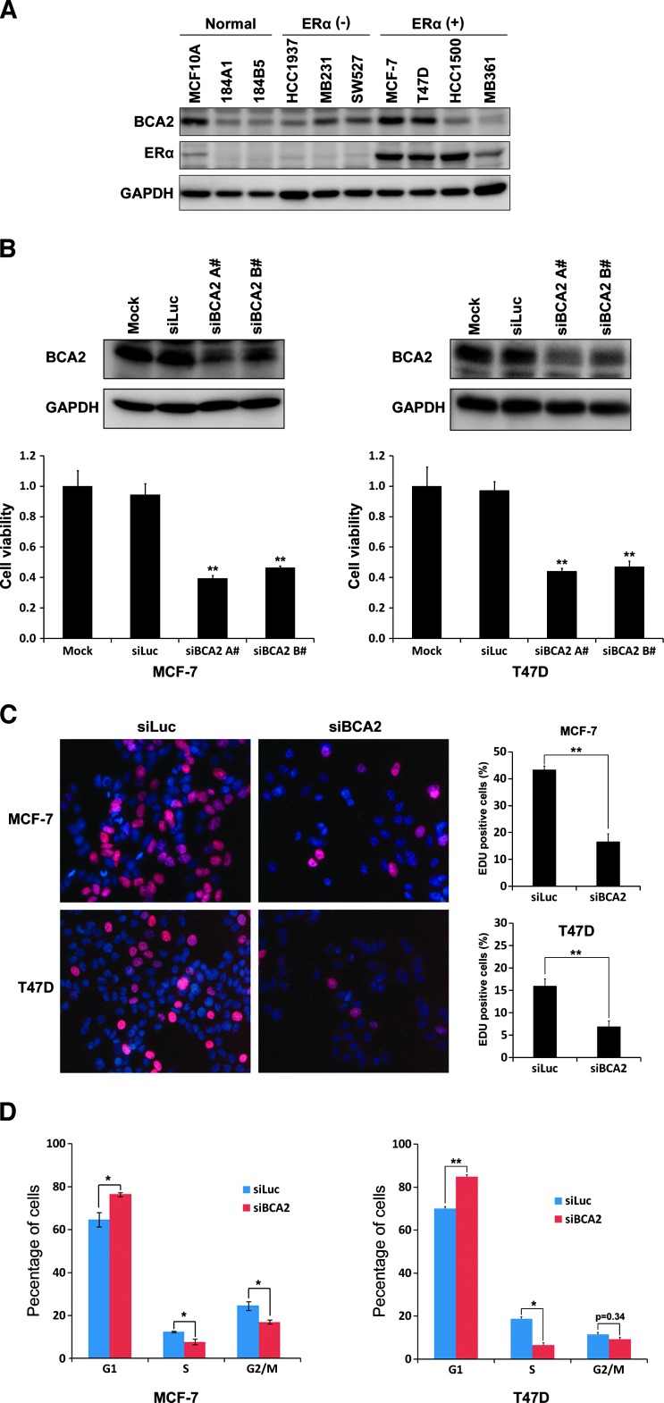
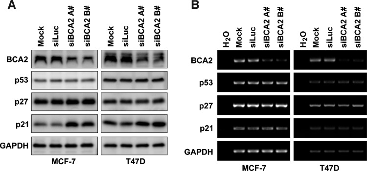
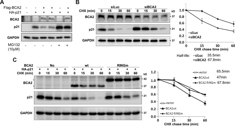
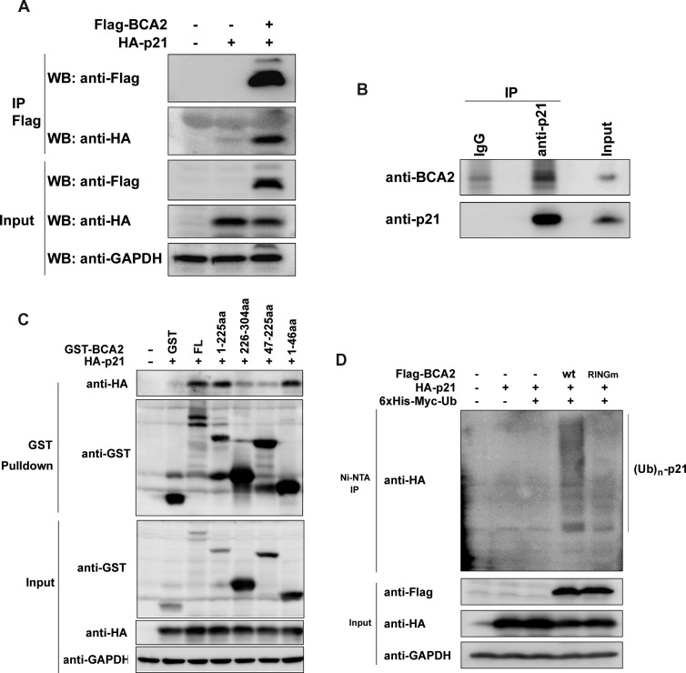
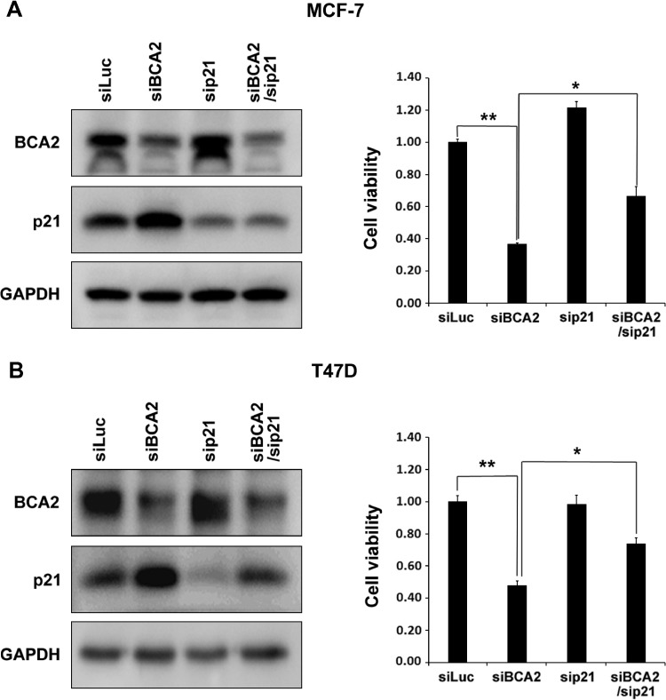
The E3 ubiquitin ligase RING finger protein 115 (RNF115), also known as breast cancer-associated gene 2 (BCA2), has previously been reported to be overexpressed in estrogen receptor α (ERα)-positive breast tumors and to promote breast cell proliferation; however, its mechanism is unknown. In this study, we demonstrated that silencing of BCA2 by small interfering RNAs (siRNAs) in two ERα-positive breast cancer cell lines, MCF-7 and T47D, decreases cell proliferation and increases the protein levels of the cyclin-dependent kinase inhibitor p21Waf/Cip1. The protein stability of p21 was negatively regulated by BCA2. BCA2 directly interacts with p21 and promotes p21 ubiquitination and proteasomal degradation. Knockdown of p21 partially rescues the cell growth arrest induced by the BCA2 siRNA. These results suggest that BCA2 promotes ERα-positive breast cancer cell proliferation at least partially through downregulating the expression of p21.


Figures

Figure 1
Figure 1
Knockdown of BCA2 inhibits breast cancer cell proliferation, DNA synthesis, and cell cycle. (A) WB analysis of BCA2 protein levels in a panel of immortalized breast epithelial cell lines and ERα-negative and ERα-positive breast cancer cell lines. GAPDH was used as a loading control. (B) MCF-7 and T47D cells were transfected for 72 hours with either Lipofectamine 2000 reagent as mock, control siLuc RNA, siBCA2-A#, or siBCA2-B#. The knockdown effect was assessed by WB analysis, and cell viability was examined using the SRB assay (**P < .01 compared to siLuc). (C) After silencing of BCA2 by siBCA2-A#, MCF-7 and T47D cells were pulse labeled with EdU Alexa Fluor 647 for 4 hours and were stained the nuclei with 10 µg/ml PI. The percentage of positive cells incorporated with EdU were shown in the histograms (right side, **P < .01). All quantitative values were from triplicate experiments. (D) After silencing of BCA2 by siBCA2-A#, MCF-7 and T47D cells were harvested, fixed, stained with PI solution, and analyzed by flow cytometry. The percentage of arrest G1 phase cells was shown in the histograms (*P < .05 and **P < .01).
Figure 2
Figure 2
BCA2 depletion upregulates the p21 expression at the protein level but not the mRNA level. (A) MCF-7 and T47D cells were transfected with either siLuc or siBCA2 for 72 hours. Protein levels of BCA2, p21, p27, and p53 were analyzed by WB analysis. (B) The mRNA levels of BCA2, p21, p27, and p53 were analyzed using semiquantitative RT-PCR. GAPDH was used as a loading control.
Figure 3
Figure 3
(A) BCA2 promotes p21 protein degradation through proteasome. Flag-BCA2 and HA-p21 were cotransfected into HEK293FT cells as indicated. The proteasome inhibitor MG132 (10 µM) was added to treat the cells for 8 hours if necessary. (B) After depletion of BCA2 by siBCA2-A# in MCF-7 cells, the cells were exposed to CHX for a time as indicated, and WB analysis was performed. The quantitative half-lives of p21 protein from two independent experiments are shown on the right side. (C) HEK293FT cells were cotransfected with HA-p21 and WT BCA2 or RING finger mutant BCA2 expression plasmids. The BCA2-RINGm protein migrates much slower than WT BCA2 possibly because of the protein structure difference under the electrophoresis condition. The cells were exposed to CHX for a time as indicated. The level of p21 protein was detected by WB analysis. The quantitative data from two independent experiments are shown on the right side.
Figure 4
Figure 4
BCA2 interacts with p21 and promotes its ubiquitination. (A) The HA-p21 expression construct was transiently cotransfected with an empty control or Flag-BCA2 expression plasmid into HEK293FT cells, respectively. After culturing for 48 hours, the cells were exposed to 10 µM MG132 for 4 hours and collected for immunoprecipitation experiments using anti-Flag M2 affinity gel. The samples were subjected to WB analysis using anti-Flag and anti-HA antibodies. Total cell lysates were used as the input control. (B) Cell lysates from exponentially growing MCF-7 cells were immunoprecipitated with the anti-p21 antibody or the rabbit IgG control. Precipitates were subjected to WB analysis with anti-p21 and anti-BCA2 antibodies. (C) The HA-p21 expression construct was transiently cotransfected with a pEBG vector as a control, PEBG-BCA2 as a positive control, or truncated GST-BCA2 expression plasmids in HEK293FT cells, respectively. Total cell lysates were collected 48 hours later and precipitated with Glutathione Sepharose 4B. The precipitated proteins were subjected to WB analysis using anti-HA and anti-GST antibodies. Total cell lysates were used as the input control. (D) HEK293FT cells were transiently cotransfected with a panel of expression plasmids as indicated. Forty-eight hours later, the cells were treated for 4 hours with 10 µM MG132 and lysed. The samples were precipitated with Ni-NTA agarose beads under a denaturing condition. The proteins were subjected to WB analysis with anti-HA and anti-Flag antibodies.
Figure 5
Figure 5
BCA2 promotes breast cancer cell proliferation partially through downregulating the expression of p21. MCF-7 and T47D cells were transiently transfected with siLuc, sip21, and siBCA2 siRNA, as indicated. Cell lysates were collected 72 hours later and subjected to WB analysis. Protein levels of p21 and BCA2 in MCF-7 (A) or T47D (B) were shown. The cell viability was measured by the SRB assay and shown in the graphs, respectively, (A) MCF-7 or (B) T47D (**P < .01).

References

    1. Carrano AC, Eytan E, Hershko A, Pagano M. SKP2 is required for ubiquitin-mediated degradation of the CDK inhibitor p27. Nat Cell Biol. 1999;1:193–199. - PubMed
    1. Yu ZK, Gervais JL, Zhang H. Human CUL-1 associates with the SKP1/SKP2 complex and regulates p21CIP1/WAF1 and cyclin D proteins. Proc Natl Acad Sci USA. 1998;95:11324–11329. - PMC - PubMed
    1. Koepp DM, Schaefer LK, Ye X, Keyomarsi K, Chu C, Harper JW, Elledge SJ. Phosphorylation-dependent ubiquitination of cyclin E by the SCFFbw7 ubiquitin ligase. Science. 2001;294:173–177. - PubMed
    1. Strohmaier H, Spruck CH, Kaiser P, Won KA, Sangfelt O, Reed SI. Human F-box protein hCdc4 targets cyclin E for proteolysis and is mutated in a breast cancer cell line. Nature. 2001;413:316–322. - PubMed
    1. Yada M, Hatakeyama S, Kamura T, Nishiyama M, Tsunematsu R, Imaki H, Ishida N, Okumura F, Nakayama K, Nakayama KI. Phosphorylation-dependent degradation of c-Myc is mediated by the F-box protein Fbw7. EMBO J. 2004;23:2116–2125. - PMC - PubMed

Publication types

MeSH terms