

Dose Reduction Technique Using a Combination of a Region of Interest (ROI) Material X-Ray Attenuator and Spatially Different Temporal Filtering for Fluoroscopic Interventions
- PMID: 24027617
- PMCID: PMC3766980
- DOI: 10.1117/12.910945
Dose Reduction Technique Using a Combination of a Region of Interest (ROI) Material X-Ray Attenuator and Spatially Different Temporal Filtering for Fluoroscopic Interventions
Abstract
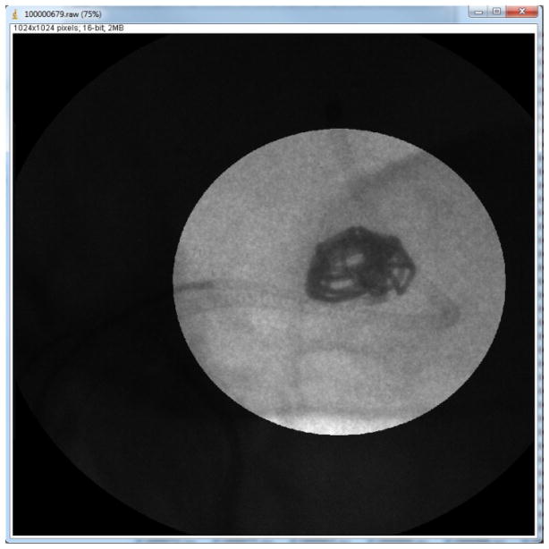
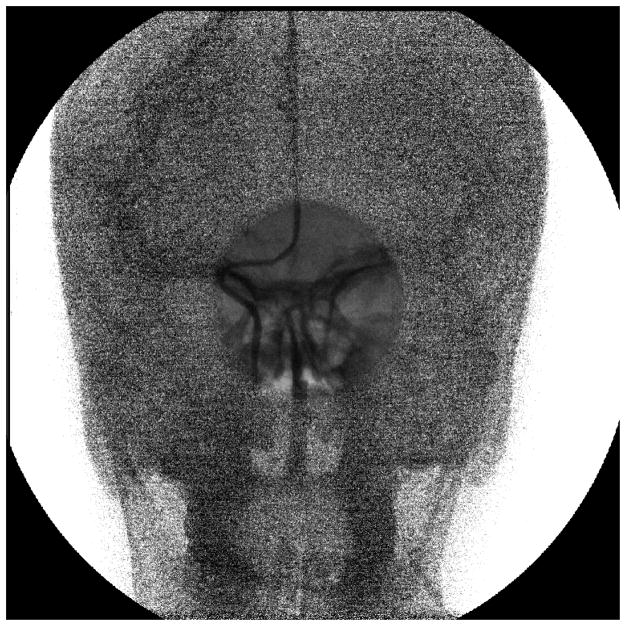
We demonstrate a novel approach for achieving patient dose savings during image-guided neurovascular interventions, involving a combination of a material x-ray region of interest (ROI) attenuator and a spatially different ROI temporal filtering technique. The part of the image under the attenuator is reduced in dose but noisy and less bright due to fewer x-ray quanta reaching the detector, as compared to the non-attenuating (or less attenuating) region. First the brightness is equalized throughout the image by post processing and then a temporal filter with higher weights is applied to the high attenuating region to reduce the noise, at the cost of increased lag; however, in the regions where less attenuation is present, a lower temporal weight is needed and is applied to preserve temporal resolution. A simulation of the technique is first presented on an actual image sequence obtained from an endovascular image guided interventional (EIGI) procedure. Then the actual implementation of the technique with a physical ROI attenuator is presented. Quantitative analysis including noise analysis and integral dose calculations are presented to validate the proposed technique.
Figures


References
-
- Kotre CJ, Marshall NW, Faulkner K. Radiation Protection Dosimetry. 3–4. Vol. 57. Nuclear Technology Publishing; 1995. Contrast -Detail Testing Techniques for Modern X Ray Image Intensifier Systems; pp. 245–247.
-
- Kotre CJ, Guibelalde E. Optimization of variable temporal averaging in digital fluoroscopy. The British Journal of Radiology. 2004;77:675–678. - PubMed
-
- Rudin S, Bednarek DR. Region of interest fluoroscopy. Med Physics. 1992 Sep-Oct;19(5):1183–1189. - PubMed
-
- Rudin S, Guterman LR, Granger WE, Bednarek DR, Hopkins LN. Application of region-of-interest imaging techniques to neurointerventional radiology. Radiology. 1996 Jun;199:870–873. - PubMed
Grants and funding
LinkOut - more resources
Full Text Sources