Multiple cementoblastoma: a rare case report
- PMID: 24027644
- PMCID: PMC3763579
- DOI: 10.1155/2013/828373
Multiple cementoblastoma: a rare case report
Abstract
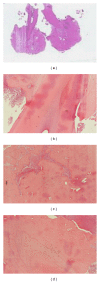
Benign cementoblastoma is a rare ectomesenchymal odontogenic tumor that originates from the root of the tooth and that is characterized by the formation of cementum-like tissue. A 60-year old man was referred to us complaining of pain in his right jaw. The patient underwent TC dental scan of the mandible, which highlighted the presence of three well-circumscribed, round, unilocular neoformations of radiopaque appearance with a radiotransparent edge, one of which was in close contact with the roots of the lower right second molar. Microscopic examination of the greater sample consisted, in its central portion, of dense mineralized acellular trabeculae of basophilic tissue cement-like, devoid of vessels, adhering to the root of the tooth, while peripherally was observed a zone of vascularized osteoid surrounded, occasionally, by a thin rim of cementoblasts mixed with fibrous tissue and inflammatory elements. This lesion was diagnosed as cementoblastoma. The second lesion appeared radiologically and histologically entirely identical to cementoblastoma, but it did not show the intimate association with the root of involved tooth. After a careful review of the literature, the diagnosis of residual cementoblastoma was made. The clinicopathologic features, treatment, and prognosis of this rare tumor are here discussed for the young dental practitioner.
Figures


References
-
- Barnes L, Eveson JW, Reichart P, Sidransky D. Pathology & Genetics Head and Neck Tumours WHO Classification of Tumours. 2005.
-
- Cawason RA, Binnie WH, Speight PM, Barrett AW, Wright JM. Lucas's Pathology of Tumors of the Oral Tissues. 5th edition 1998.
-
- Piattelli A, Di Alberti L, Scarano A, Piattelli M. Benign cementoblastoma associated with an unerupted third molar. Oral Oncology. 1998;34(3):229–231. - PubMed
-
- Baart JA, Lekkas C, Van der Waal I. Residual cementoblastoma of the mandible. Journal of Oral Pathology and Medicine. 1991;20(6):300–302. - PubMed
-
- Pacifici L, Tallarico M, Bartoli A, Ripari A, Cicconetti A. Benign cementoblastoma: a clinical case of conservative surgical treatment of the involved tooth. Minerva Stomatologica. 2004;53(11-12):685–691. - PubMed
LinkOut - more resources
Full Text Sources
Other Literature Sources

