. 2013 Dec:250:143-50.
doi: 10.1016/j.expneurol.2013.09.001. Epub 2013 Sep 9.

Presenilins regulate the cellular activity of ryanodine receptors differentially through isotype-specific N-terminal cysteines

Affiliations

Presenilins regulate the cellular activity of ryanodine receptors differentially through isotype-specific N-terminal cysteines

Andrew J Payne et al. Exp Neurol. 2013 Dec.

Abstract

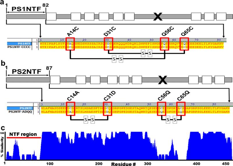
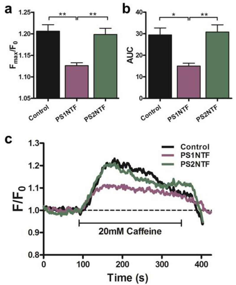
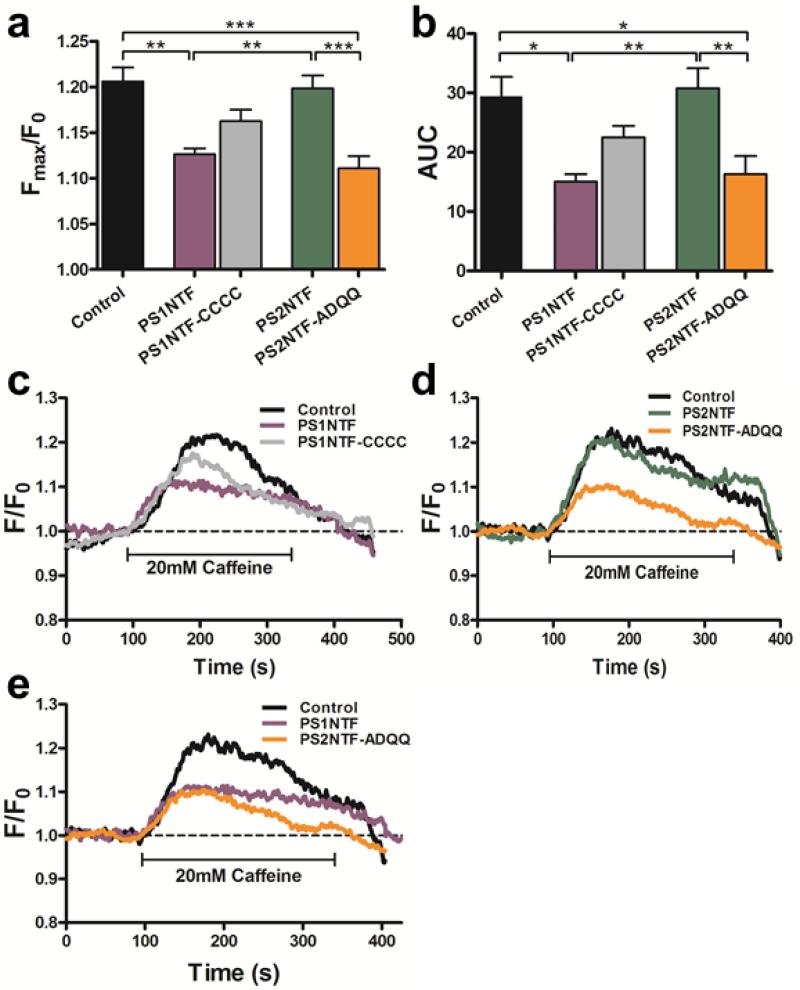
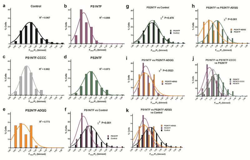
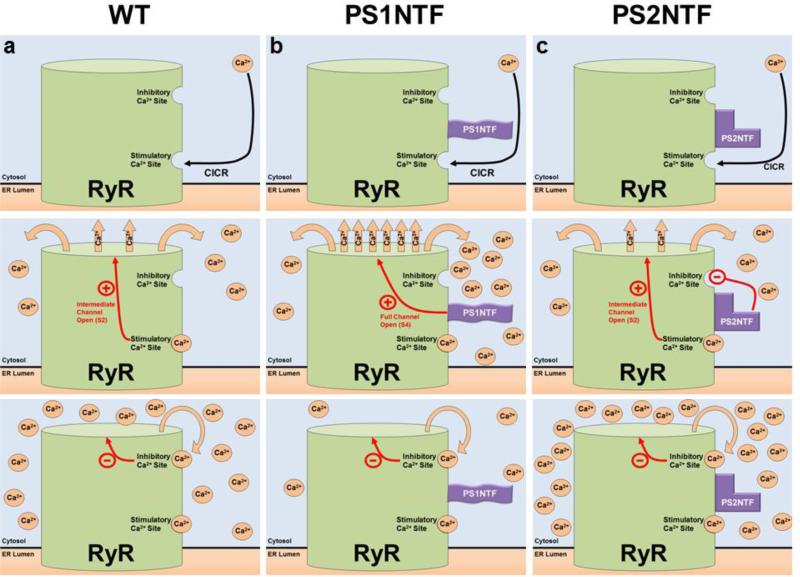
Presenilins (PS), endoplasmic reticulum (ER) transmembrane proteins, form the catalytic core of γ-secretase, an amyloid precursor protein processing enzyme. Mutations in PS lead to Alzheimer's disease (AD) by altering γ-secretase activity to generate pathologic amyloid beta and amyloid plaques in the brain. Here, we identified a novel mechanism where binding of a soluble, cytosolic N-terminal domain fragment (NTF) of PS to intracellular Ca(2+) release channels, ryanodine receptors (RyR), controls Ca(2+) release from the ER. While PS1NTF decreased total RyR-mediated Ca(2+) release, PS2NTF had no effect at physiological Ca(2+) concentrations. This differential function and isotype-specificity is due to four cysteines absent in PS1NTF, present, however, in PS2NTF. Site-directed mutagenesis targeting these cysteines converted PS1NTF to PS2NTF function and vice versa, indicating differential RyR binding. This novel mechanism of intracellular Ca(2+) regulation through the PS-RyR interaction represents a novel target for AD drug development and the treatment of other neurodegenerative disorders that critically depend on RyR and PS signaling.

Keywords: Alzheimer's disease; Endoplasmic reticulum; Intracellular calcium; Neuroprotection; Oxidative stress.


Figures

Figure 1
Figure 1. Alignment and predicted disulfide bridges of PS1NTF and PS2NTF and their mutants
Cysteine sites of PS2NTF were mutated to the complementary residues of PS1NTF and vice versa, resulting in the PS2NTF-ADQQ and PS1NTF-CCCC constructs, respectively. A) Schematic representation of PS1. The soluble N-terminus fragment used in this study is indicated as PS1NTF (white rectangles, TM domains; “x”, endogenous cleavage site). Red boxes indicate mutagenesis sites, where the ADQQ residues of PS1NTF were replaced with complementary cysteine residues of PS2 resulting in the mutant PS1NTF-CCCC. Thick black lines between red boxes represent predicted disulfide bridges (http://scratch.proteomics.ics.uci.edu/; 3/6/2013). B) Schematic representation of PS2 (figure markings the same as 1a). Red boxes indicate mutagenesis sites where cysteine residues of PS2NTF were replaced with complementary residues of PS1 resulting in the mutant PS2NTF-ADQQ (black lines represent predicted disulfide bridges). C) The homology alignment for PS1 and PS2 identifies regions of high variability at the cytosolic N-termini (residues 1-80, blue tracing is proportional to the percent homology between PS1 and PS2) and endogenous cleavage regions (residues #320-370). This contrasts with the high homology of the proteins overall. The different effects of PS1 and PS2 are likely caused by the sequence differences of these two regions between the proteins.
Figure 2
Figure 2. Pharmacologically elicited intracellular calcium release from RyR is significantly attenuated by PS1NTF but not PS2NTF
A) Cytoplasmic expression of PS1NTF reduced the maximum amplitude (Fmax/F0) of the RyR mediated calcium transient after pharmacological stimulation of SH-SY5Y cells with 20mM caffeine (n=8), however, cytoplasmic expression of PS2NTF (n=11) has no effect when compared to untransfected control (n=8). B) AUC of calcium transients measured after pharmacological stimulation as a measure of total intracellular calcium release was significantly reduced in cells expressing PS1NTF but remained unaltered in PS2NTF expressing cells (n= 8 and 11, respectively). Data is shown as mean ± SEM with significance determined by one-way ANOVA and Bonferroni's post-hoc test. [* P <0.05; ** P <0.01]. C) Representative traces of calcium transients after pharmacological stimulation of SH-SY5Y cells expressing cytoplasmic PS1NTF or PS2NTF.
Figure 3
Figure 3. Mutation of PS2NTF cysteines to the complementary PS1NTF residues exchanges ability to regulate RyR calcium release
A) The amplitude of RyR-mediated, pharmacologically elicited intracellular calcium release in SH-SY5Y cells was measured as the maximum emission fluorescence value over baseline (Fmax/F0). Cells expressing PS2NTF-ADQQ (amino acids 1-87 of PS2 with the mutations C14A, C31D, C56Q, C65Q; n=8) showed similar attenuation of maximal calcium release (Fmax/F0) as cells expressing PS1NTF (n=8). Mutation of PS1NTF to PS1NTF-CCCC (amino acids 1-82 of PS1 with the mutations A14C, D31C, Q56C, Q65C; n=8) attenuated the PS1NTF effect on RyR calcium release but did not fully restore Fmax/F0 values measured in PS2NTF expressing (n=11) or control cells (n=8) resulting in a distinct intermediate response that was not statistically different from either the PS1NTF or PS2NTF expressing cells. B) AUC of calcium transients measured after pharmacological stimulation of SH-SY5Y cells expressing the recombinant PS-NTF domains mirror the results seen for the Fmax/F0 values. Data is shown as mean ± SEM; asterisks indicate significance by one-way ANOVA with Bonferroni's post-hoc test [* P <0.05; ** P <0.01; *** P <0.001]. Representative traces of calcium transients produced by pharmacological stimulation of SH-SY5Y cells expressing PS1NTF or PS1NTF-CCCC (C) and PS2NTF or PS2NTF-ADQQ (D). Representative traces show the similar lowering effect of PS1NTF and the mutant PS2NTF-ADQQ on RyR calcium release (E).
Figure 4
Figure 4. Amplitudes of the RyR mediated calcium transients binned at 5% intervals are Gaussian normally distributed. PS2NTF-ADQQ and PS1NTF shift the distribution curve left, indicating attenuation of RyR-mediated intracellular calcium release
Fmax/F0 values per cell were binned at 5% width (abscissae) as a percentage of the total number of Fmax/F0 values (ordinates) collected for each experimental condition. Columns indicate the percentage of Fmax/F0 values that fell between the bin indicated on the abscissa (α) but below the next higher value on the abscissa such that α < × < (α+0.05). Lines are the non-linear regression to determine the Gaussian distribution (1,000 iterations) with the corresponding R2 value indicated. All treatment groups were found to be normally distributed. Data and analyses for control SH-SY5Y cells (A), PS1NTF (B), PS1NTF-CCCC (C), PS2NTF (D), and PS2NTF-ADQQ (E). Binned histograms were compared by the stringent Pearson's chi-squared (χ2) test and key comparisons are shown (F-K). Both PS1NTF (F, I, J) and PS2NTF-ADQQ (H, I, K) significantly shift the Fmax/F0 distribution leftward, indicating attenuation of RyR calcium release vs. control. PS2NTF (G) was not different from controls while PS1NTF-CCCC (J) resulted in an intermediate effect. Pearson's χ2 test results not indicated on the graphs above: J) PS1NTF / PS2NTF, χ2 P<0.001; PS1NTF / PS1NTF-CCCC, χ2 P<0.05; PS2NTF / PS1NTF-CCCC, χ2 P<0.05. K) Control / PS1NTF, χ2 P<0.001; Control / PS2NTF-ADQQ, χ2 P<0.001; PS1NTF / PS2NTF-ADQQ, χ2 n.s. Pearson's χ2 tests for all treatment groups are given in Supplementary Data Table 4.
Figure 5
Figure 5. Proposed mechanism of RyR calcium release regulated by the N-termini of PS1 and PS2
PS1NTF and PS2NTF have distinct effects on RyR calcium induced calcium release (CICR) from ER stores. A) The mechanism of RyR regulation by Ca2+ binding at the high affinity stimulatory site results in moderate channel opening. Ca2+ is released until the local Ca2+ concentration rises to the point at which the low affinity, inhibitory Ca2+ binding site is occupied resulting in closure of the channel. B) The N-terminus of PS1 (PS1NTF) binds RyR and increases calcium release from the ER. Occupation of the RyR high affinity stimulatory site by Ca2+ causes channel gating and Ca2+ release. Bound PS1NTF increases channel opening, causing rapid RyR Ca2+ release until the low affinity inhibitory Ca2+ site is bound, closing the channel and terminating ER Ca2+ release. The increased rate of Ca2+ release due to PS1NTF binding results in overall reduced Ca2+ release because inhibitory concentrations are reached in less time. C) The N-terminus of PS2 (PS2NTF) binds to RyR. Ca2+ ion binding at the high affinity stimulatory site of RyR causes channel gating and Ca2+ release. PS2NTF has no effect on channel gating but blocks Ca2+ inhibition of the RyR channel at high cytosolic calcium concentrations. Significantly elevated cytosolic Ca2+ concentrations result in binding of Ca2+ at the low affinity inhibitory site eventually closing the channel and ending calcium release, however, resulting in an overall higher cytosolic Ca2+ concentration.

References

    1. Alzheimer's Association 2012 Alzheimer's disease facts and figures. Alzheimers Dement. 2012;8:131–168. - PubMed
    1. Alzheimer's Disease Collaborative Group The structure of the presenilin 1 (S182) gene and identification of six novel mutations in early onset AD families. Alzheimer's Disease Collaborative Group. Nat Genet. 1995;11:219–222. - PubMed
    1. Borchelt DR, Thinakaran G, Eckman CB, Lee MK, Davenport F, Ratovitsky T, Prada CM, Kim G, Seekins S, Yager D, Slunt HH, Wang R, Seeger M, Levey AI, Gandy SE, Copeland NG, Jenkins NA, Price DL, Younkin SG, Sisodia SS. Familial Alzheimer's disease-linked presenilin 1 variants elevate Abeta1-42/1-40 ratio in vitro and in vivo. Neuron. 1996;17:1005–1013. - PubMed
    1. Burroughs SL, Duncan RS, Rayudu P, Kandula P, Payne AJ, Clark JL, Koulen P, Kaja S. Plate reader-based assays for measuring cell viability, neuroprotection and calcium in primary neuronal cultures. J Neurosci Methods. 2012;203:141–145. - PMC - PubMed
    1. Borghi R, Piccini A, Barini E, Cirmena G, Guglielmotto M, Tamagno E, Fornaro M, Perry G, Smith MA, Garuti A, Tabaton M. Upregulation of presenilin 1 in brains of sporadic, late-onset Alzheimer's disease. J Alzheimers Dis. 2010;22(3):771–775. - PubMed

Publication types

LinkOut - more resources