Comparative Study
. 2013 Sep;6(9):973-83.
doi: 10.1016/j.jcmg.2013.04.012.

Libman-Sacks endocarditis and embolic cerebrovascular disease

Affiliations
Comparative Study

Libman-Sacks endocarditis and embolic cerebrovascular disease

Carlos A Roldan et al. JACC Cardiovasc Imaging. 2013 Sep.

Abstract

Objectives: The aim of this study was to determine whether Libman-Sacks endocarditis is a pathogenic factor for cerebrovascular disease (CVD) in systemic lupus erythematosus (SLE).

Background: A cardioembolic pathogenesis of SLE CVD manifested as: 1) neuropsychiatric systemic lupus erythematosus (NPSLE), including stroke and transient ischemic attacks (TIA); 2) neurocognitive dysfunction; and 3) magnetic resonance imaging of focal brain lesions has not been established.

Methods: A 6-year study of 30 patients with acute NPSLE (27 women, 38 ± 12 years of age), 46 age- and sex-matched SLE controls without NPSLE (42 women, 36 ± 12 years of age), and 26 age- and sex-matched healthy controls (22 women, 34 ± 11 years of age) who underwent clinical and laboratory evaluations, transesophageal echocardiography, carotid duplex ultrasound, transcranial Doppler ultrasound, neurocognitive testing, and brain magnetic resonance imaging/magnetic resonance angiography. Patients with NPSLE were re-evaluated after 4.5 months of therapy. All patients were followed clinically for a median of 52 months.

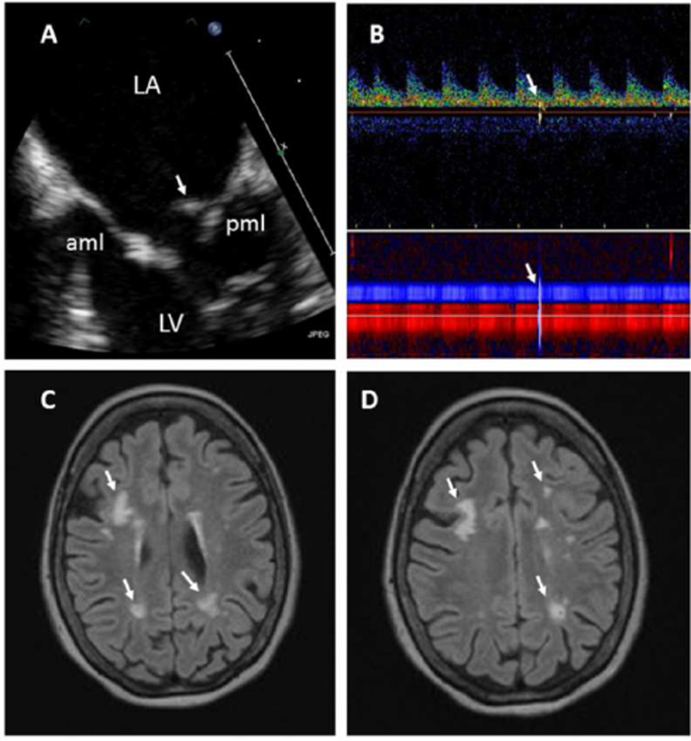
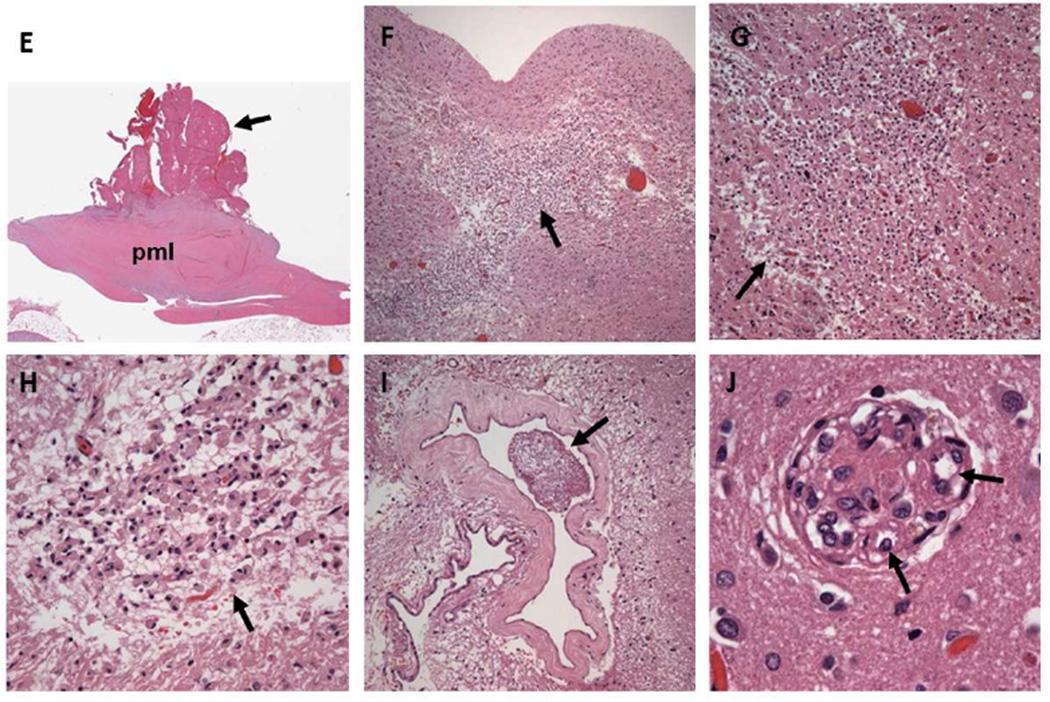
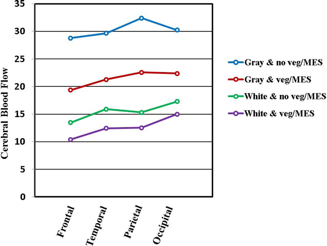
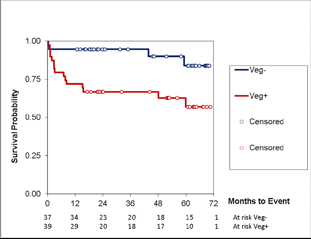
Results: Libman-Sacks vegetations (87%), cerebromicroembolism (27% with 2.5 times more events per hour), neurocognitive dysfunction (60%), and cerebral infarcts (47%) were more common in NPSLE than in SLE (28%, 20%, 33%, and 0%) and healthy controls (8%, 0%, 4%, and 0%, respectively) (all p ≤ 0.009). Patients with vegetations had 3 times more cerebromicroemboli per hour, lower cerebral blood flow, more strokes/TIA and overall NPSLE events, neurocognitive dysfunction, cerebral infarcts, and brain lesion load than those without (all p ≤ 0.01). Libman-Sacks vegetations were independent risk factors of NPSLE (odds ratio [OR]: 13.4; p < 0.001), neurocognitive dysfunction (OR: 8.0; p = 0.01), brain lesions (OR: 5.6; p = 0.004), and all 3 outcomes combined (OR: 7.5; p < 0.001). Follow-up re-evaluations in 18 of 23 (78%) surviving patients with NPSLE demonstrated improvement of vegetations, microembolism, brain perfusion, neurocognitive dysfunction, and lesion load (all p ≤ 0.04). Finally, patients with vegetations had reduced event-free survival time to stroke/TIA, cognitive disability, or death (p = 0.007).

Conclusions: The presence of Libman-Sacks endocarditis in patients with SLE was associated with a higher risk for embolic CVD. This suggests that Libman-Sacks endocarditis may be a source of cerebral emboli.

Keywords: CVD; Libman-Sacks endocarditis; MRA; NPSLE; SLE; TEE; TIA; TTE; cerebrovascular disease; magnetic resonance angiography; microembolism; neuropsychiatric systemic lupus erythematosus; stroke; systemic lupus erythematosus; transesophageal echocardiography; transient ischemic attack; transthoracic echocardiography.


Figures

Figure 1
Figure 1. Part 1. 55 year old female with SLE and acute transient ischemic attack
A. This TEE view demonstrates a moderate size, elongated, sessile, and heterogeneously echoreflectant Libman-Sacks vegetation (arrow) on the atrial side of the posterior mitral leaflet (pml) (video clip Figure 1A). Moderate thickening and sclerosis with decreased mobility of the mid and distal portions of the anterior (aml) and posterior (pml) mitral leaflets is noted. B. Transcranial Doppler demonstrates a microembolic signal on the spectral Doppler (upper arrow) and vessels lumen (lower arrow) traveling through the left middle cerebral artery (red power M-mode) and anterior communicating artery (blue power M-mode). C,D. Brain MRI demonstrates multiple, bilateral, and variable size periventricular and deep white matter infarcts (arrows). This patient had 75 brain lesions and a lesion load of 6.04 cm3. Her global neurocognitive z-score was −4.28 indicative of severe neurocognitive dysfunction. Abbreviations: LA = left atrium, LV = left ventricle. Part 2. 55 year old female with SLE and acute transient ischemic attack who during follow up died from a large intracerebral hemorrhage. Histopathology with H&E stains demonstrates: E (H&E 40X): Thickening and fibrosis of the posterior mitral leaflet (pml) with a well adhered, verrucoid, fibrinous vegetation (arrow). F (H&E 20X): Subacute cerebral infarct at the junction of the white and gray matter with necrotic debris and moderate cellular infiltration (arrow). G,H (H&E 40X, 100X): Old deeper (white matter) infarct with liquefactive necrosis, residual macrophages, and gliosis (arrows). I (H&E 100X): Large cerebral vessel with fibrin thrombi (arrow). J (H&E 100X): Small cerebral vessel with fibrin thrombi and neoangiogenesis (arrows). Multiple subacute and old microinfarcts and fibrin thrombosed microvasculature with neoangiogenesis characteristic of chronic cardiothromboembolic disease were demonstrated in both cerebral hemispheres.
Figure 2
Figure 2. Cerebral blood flow in SLE patients with vegetations and cerebromicroembolism
Cerebral blood flow (ml/min/100 grams of tissue) in the gray and white matter is significantly lower in 11 patients with vegetations and cerebromicroembolism as compared to 24 patients with neither across the 4 cerebral lobes and left and right hemispheres (RM ANOVA p≤0.001 for both gray and white matter). Abbreviations: Gray & no veg/MES, White & no veg/MES = gray or white matter perfusion in patients with no vegetations and no cerebromicroembolism; Gray & veg/MES, White & veg/MES = gray or white matter perfusion in patients with vegetations and cerebromicroembolism.
Figure 3
Figure 3. Kaplan-Meier event free survival in SLE patients with and without vegetations
During follow-up, the event free survival from stroke/TIA, cognitive disability, or death of patients with vegetations (Veg+) was significantly lower than in those without vegetations (Veg−) (p=0.007).

Comment in

References

    1. Hanly JG, Urowitz MB, Su L, et al. Prospective analysis of neuropsychiatric events in an international disease inception cohort of patients with systemic lupus erythematosus. Ann Rheum Dis. 2010;69:529–535. - PMC - PubMed
    1. The American College of Rheumatology nomenclature and case definitions for neropsychiatric lupus erythematosus. Arthritis Rheum. 1999;42:599–608. - PubMed
    1. Luyendijk J, Steens SC, Ouwendijk WJ, et al. Neuropsychiatric systemic lupus erythematosus: lessons learned from magnetic resonance imaging. Arthritis Rheum. 2011;63:722–732. - PubMed
    1. Sibbitt WL, Jr, Schmidt PJ, Hart BL, Brooks WM. Fluid Attenuated Inversion Recovery (FLAIR) imaging in neuropsychiatric systemic lupus erythematosus. J Rheumatol. 2003;30:1983–1989. - PubMed
    1. Bernatsky S, Clarke A, Gladman DD, et al. Mortality related to cerebrovascular disease in systemic lupus erythematosus. Lupus. 2006;15:835–839. - PubMed

Publication types