Review
. 2013 Oct;38(10):494-506.
doi: 10.1016/j.tibs.2013.07.004. Epub 2013 Sep 10.

Stress granules and cell signaling: more than just a passing phase?

Affiliations
Review

Stress granules and cell signaling: more than just a passing phase?

Nancy Kedersha et al. Trends Biochem Sci. 2013 Oct.

Abstract

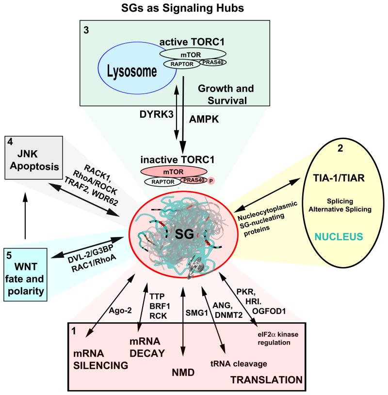
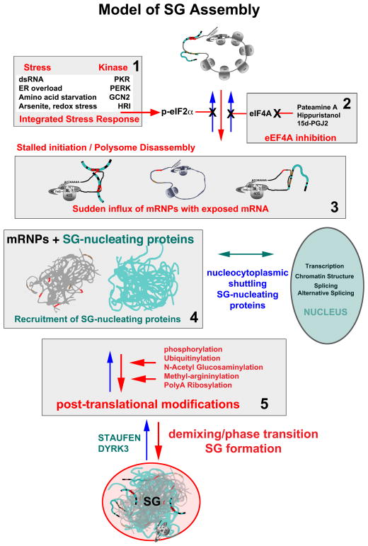
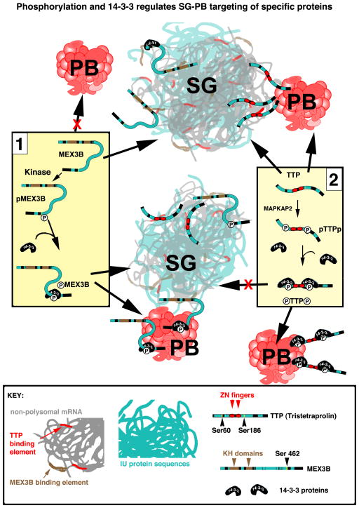
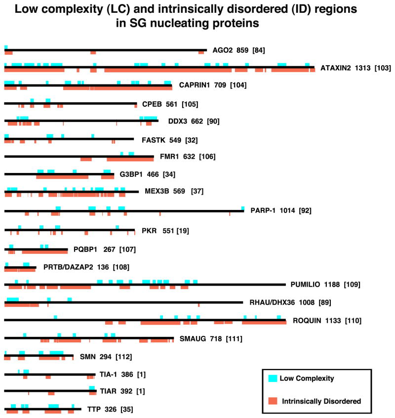
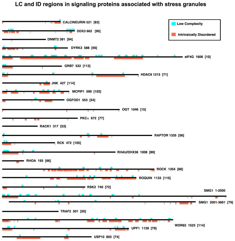
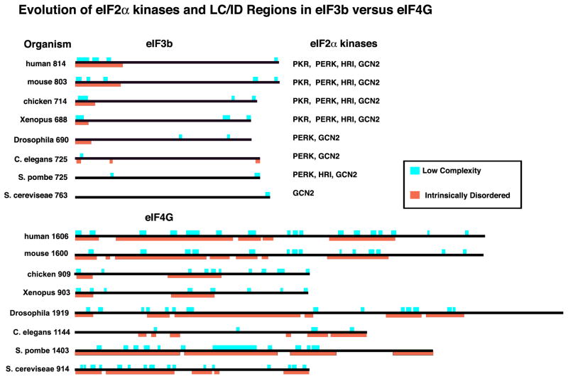
Stress granules (SGs) contain translationally-stalled mRNAs, associated preinitiation factors, and specific RNA-binding proteins. In addition, many signaling proteins are recruited to SGs and/or influence their assembly, which is transient, lasting only until the cells adapt to stress or die. Beyond their role as mRNA triage centers, we posit that SGs constitute RNA-centric signaling hubs analogous to classical multiprotein signaling domains such as transmembrane receptor complexes. As signaling centers, SG formation communicates a 'state of emergency', and their transient existence alters multiple signaling pathways by intercepting and sequestering signaling components. SG assembly and downstream signaling functions may require a cytosolic phase transition facilitated by intrinsically disordered, aggregation-prone protein regions shared by RNA-binding and signaling proteins.

Keywords: cell signaling; intrinsically disordered; protein aggregation; stress granules; translation.


Figures

Figure I
Figure I. Evolution of eIF2α kinases and LC/ID regions in eIF3b versus eIF4G
Proteins are shown to scale, with LC (aqua) and ID (orange) regions and total number of amino acids as indicated. The evolution of an increasingly large LC/ID region at the N-terminus of eIF3b parallels the appearance of PKR, the importance of phospho-eIF2α in SG assembly, and of the inclusion of eIF3 in SGs.
Fig. 1
Fig. 1. Model of SG Assembly
Stress induced phosphorylation of eIF2α (panel 1) or inactivation of eIF4A (panel 2) slows initiation rates and results in a sudden increase in non-polysomal mRNPs (panel 3), containing exposed mRNA regions previously masked by translating ribosomes. This transiently “naked” mRNA immediately binds any available mRNA binding proteins (panel 3: blue noodles), many of which are LC/ID rich (see Fig. 3), and many of which normally shuttle between the cytoplasm and the nucleus. This mRNA/protein mix is highly dynamic as the various mRNA binding proteins “trade up” to find their optimal binding sequences. The protein-mRNA mix “condenses” into immiscible droplets (panel 4), and the phase transition appears to be promoted by post-translational modifications of SG-associated proteins (panel 5). Once formed, SGs become hubs that intercept and interact with proteins involved in multiple signaling pathways, mRNA functions, and nuclear functions to influence global cell processes. Note that many of the steps shows here happen concurrently rather than in a linear sequence.
Fig. 2
Fig. 2. Phosphorylation and 14-3-3 regulates SG-PB targeting of specific proteins
MEX3B (Panel 1) possesses two KH domains (brown) and considerable ID sequence (aqua, also see Fig. 3). Non-phosphorylated MEX3B associates with SGs but not PBs. Phospho-MEX3B associates with both SGs and promotes SG-PB fusion . Its interaction with PBs requires phosphorylation of serine 462 and 14-3-3 binding, which are not required for its targeting to SGs . In contrast (Panel 2), non-phosphorylated TTP binds mRNA through its zinc fingers (red), associates with both SGs and PBs, and tethers SG and PB together , . MAPKAP2-induced phosphorylation of serines 60 and 186 promotes 14-3-3 binding, removing TTP from SGs while allowing its continued association with PBs , leading to the separation of SGs from PBs. The binding of 14-3-3 proteins to ID regions (proteins shown to scale) may stabilize the disordered regions, “freezing” them into a locked conformation (represented here by a rigid linear shape), thus taking TTP and perhaps other individual SG-associated proteins “out of phase” with the rapid and fleeting interactions within SGs.
Figure 3
Figure 3. Low complexity (LC) and intrinsically disordered (ID) regions in SG nucleating proteins
SG nucleating proteins are shown to scale and their LC (aqua) and ID (orange) regions are indicated. The numbers to the right of each schematic indicates the number of amino acids in the isoform shown (usually the largest one); numbers in brackets indicate the reference. LC (aqua) regions were obtained from the NCBI “conserved domains” graphic, which calculates low complexity regions using the SEG program . Intrinsically Disordered regions were determined using the programs on the ANCHOR website (http://anchor.enzim.hu/), in which regions of disorder exceeding 50% on the Intrinsically Unordered histograms were graphically rendered (orange). Note that all SG-nucleating proteins contain at least some LC/ID regions, and that some SG-nucleating signaling proteins (OGFOD1, DYRK3) are shown in Fig. 4 rather than here.
Figure 4
Figure 4. LC and ID regions in signaling proteins associated with stress granules
Proteins are shown to scale, with LC (aqua) and ID (orange) regions indicated. Note that several SG-associated signaling proteins (RACK1, TRAF2) are highly structured and devoid of LC/ID regions; these proteins bind to SG-integral factors (eIF3 and 40S ribosomal subunits) and proteins (eIF4G) that presumably regulates their recruitment to SGs via more canonical binding of rigid domains. Note that some proteins here (DYRK3, OGFOD1) are also SG-nucleating proteins. The numbers to the right of each schematic indicates the number of amino acids in the isoform shown (usually the largest one); numbers in brackets indicate the reference.
Figure 5
Figure 5. SGs as Signaling Hubs
SG (center) assembly and disassembly are in dynamic equilibrium with protein translation and mRNA metabolism (Panel 1); their formation is nucleated by protein effectors of mRNA silencing, decay, quality control (nonsense-mediated decay, NMD), tRNA cleavage, or translation). Nucleocytoplasmic shuttling proteins (panel 2) comprise a major class of SG-nucleating proteins such as TIA-1/TIAR, whose nuclear functions involve alternative splicing; this process may be altered when TIA-1/R are sequestered in SGs. TORC1 signaling (panel 3) both regulates and is regulated by SG assembly through DYRK3 (see text); regulation of TORC1 by AMPK may occur during cold shock . Multiple signaling proteins sequestered in SGs are effectors of JNK (panel 4) and WNT (panel 5) signaling. Double arrows signify reversible changes in location (nucleus, cytoplasm, lysosome) or state (soluble versus partitioned into SGs via a phase transition).

References

    1. Kedersha NL, Gupta M, Li W, Miller I, Anderson P. RNA-binding proteins TIA-1 and TIAR link the phosphorylation of eIF-2α to the assembly of mammalian stress granules. J Cell Biol. 1999;147:1431–41. - PMC - PubMed
    1. Anderson P, Kedersha N. Stress granules: the Tao of RNA triage. Trends in biochemical sciences. 2008;33:141–50. - PubMed
    1. Harding H, Novoa Y, Zhang H, Zeng R, Wek M, Schapira M, et al. Regulated translation initiation controls stress-induced gene expression in mammalian cells. Mol Cell. 2000;6:1099–108. - PubMed
    1. Harding HP, Zhang Y, Bertolotti A, Zeng H, Ron D. Perk is essential for translational regulation and cell survival during the unfolded protein response. Mol Cell. 2000;5:897–904. - PubMed
    1. Srivastava SP, Kumar KU, Kaufman RJ. Phosphorylation of eukaryotic translation initiation factor 2 mediates apoptosis in response to activation of the double-stranded RNA- dependent protein kinase. The Journal of biological chemistry. 1998;273:2416–23. - PubMed

LinkOut - more resources