Review
. 2013 Sep 13;113(7):922-32.
doi: 10.1161/CIRCRESAHA.113.300216.

Patching the heart: cardiac repair from within and outside

Affiliations
Review

Patching the heart: cardiac repair from within and outside

Lei Ye et al. Circ Res. .

Abstract

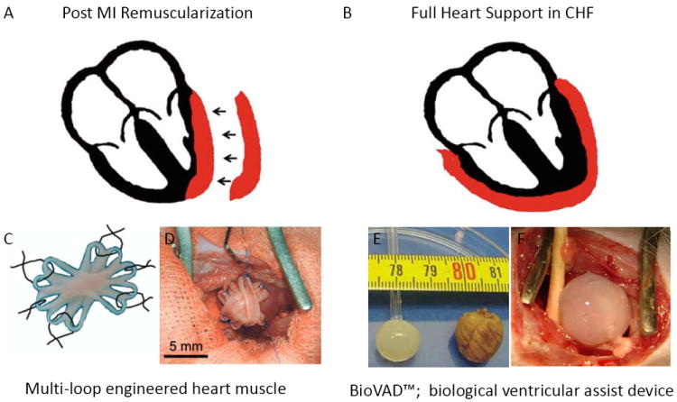
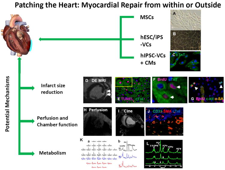
Transplantation of engineered tissue patches containing either progenitor cells or cardiomyocytes for cardiac repair is emerging as an exciting treatment option for patients with postinfarction left ventricular remodeling. The beneficial effects may evolve directly from remuscularization or indirectly through paracrine mechanisms that mobilize and activate endogenous progenitor cells to promote neovascularization and remuscularization, inhibit apoptosis, and attenuate left ventricular dilatation and disease progression. Despite encouraging results, further improvements are necessary to enhance current tissue engineering concepts and techniques and to achieve clinical impact. Herein, we review several strategies for cardiac remuscularization and paracrine support that can induce cardiac repair and attenuate left ventricular dysfunction from both within and outside the myocardium.

Keywords: heart failure; paracrine; tissue engineering; tissue therapy.


Figures

Figure 1
Figure 1
Applications of engineered heart muscle (EHM). Schematic presentation of (A) an EHM patch and (B) an EHM pouch (red). (C) Illustration of a multi-loop EHM with five loops fused together to form an asterisk-shaped stack with a solid center surrounded by 10 loops that are used for surgical fixation of the EHM graft. (D) The patch was fixed over the site of infarction in rats with six single-knot sutures. (E) The dimensions of an EHM pouch and an explanted rat heart are shown. (F) An EHM pouch was implanted over a healthy rat heart to simulate its use as a biological ventricular assist device. Panels C and D are modified from Zimmermann et al. Nat Med. 2006; 12(4): 452-8; panels E and F are modified from Yildirim et al. Circulation 2007;116(11 Suppl): I16-23.
Figure 2
Figure 2
The benefits associated with our in vivo method for cardiac patch application. A circular 3D porous biodegradable cardiac patch (blue) is created over the infarcted region by mixing thrombin and fibrinogen solutions that contain different progenitor cell types, such as mesenchymal stem cells (MSCs), vascular cells (VCs) generated through the controlled differentiation of either human embryonic stem cells (hESCs) or human induced pluripotent stem cells (hiPSCs), or both hiPSC-VCs and cardiomyocytes (hiPSC-CMs). The fibrinogen can be modified to bind peptides for different purposes, such as guiding differentiation or impeding apoptosis. The solution typically solidifies in less than one minute to form a 3D porous cardiac patch that provides structural support as well as a platform for the transplanted progenitor cells, and ultimately increases the cell engraftment rate. The transplanted cells release growth factors and other cytokines that reduce apoptosis, promote angiogenesis, and activate endogenous mechanisms for cardiomyocyte renewal, which leads to declines in infarct size and to improvements in myocardial perfusion, metabolism, and contractile function. Measurements of in vivo myocardial bioenergetics and the ATP turnover rate (via 31P magnetization saturation transfer) suggest that the patch also protects against adverse changes in cardiomyocyte energy metabolism, perhaps by reducing wall stress and bulging at the site of the infarction. Collectively, these benefits improve cardiac contractile function and impede the progression of LV dilatation. (Panels B and K courtesy of Xiong Q et al Circ Res. 2012 ;111(4):455-68; Panels F, G, I, J and L, courtesy of Xiong Q et al Circulation 2013; 127(9): 997-1008).

References

    1. McMurray JJ, Pfeffer MA. Heart failure. Lancet. 2005;365:1877–1889. - PubMed
    1. Orlic D, Kajstura J, Chimenti S, Jakoniuk I, Anderson SM, Li B, Pickel J, McKay R, Nadal-Ginard B, Bodine DM, Leri A, Anversa P. Bone marrow cells regenerate infarcted myocardium. Nature. 2001;410:701–705. - PubMed
    1. Beltrami AP, Barlucchi L, Torella D, Baker M, Limana F, Chimenti S, Kasahara H, Rota M, Musso E, Urbanek K, Leri A, Kajstura J, Nadal-Ginard B, Anversa P. Adult cardiac stem cells are multipotent and support myocardial regeneration. Cell. 2003;114:763–776. - PubMed
    1. Muller-Ehmsen J, Whittaker P, Kloner RA, Dow JS, Sakoda T, Long TI, Laird PW, Kedes L. Survival and development of neonatal rat cardiomyocytes transplanted into adult myocardium. Journal of molecular and cellular cardiology. 2002;34:107–116. - PubMed
    1. Murry CE, Whitney ML, Laflamme MA, Reinecke H, Field LJ. Cellular therapies for myocardial infarct repair. Cold Spring Harb Symp Quant Biol. 2002;67:519–526. - PubMed

Publication types