. 2013 Oct 31;122(18):3129-37.
doi: 10.1182/blood-2013-06-508432. Epub 2013 Sep 12.

Analysis of the clonal growth and differentiation dynamics of primitive barcoded human cord blood cells in NSG mice

Affiliations

Analysis of the clonal growth and differentiation dynamics of primitive barcoded human cord blood cells in NSG mice

Alice M S Cheung et al. Blood. .

Abstract

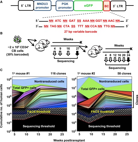
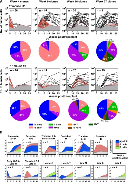
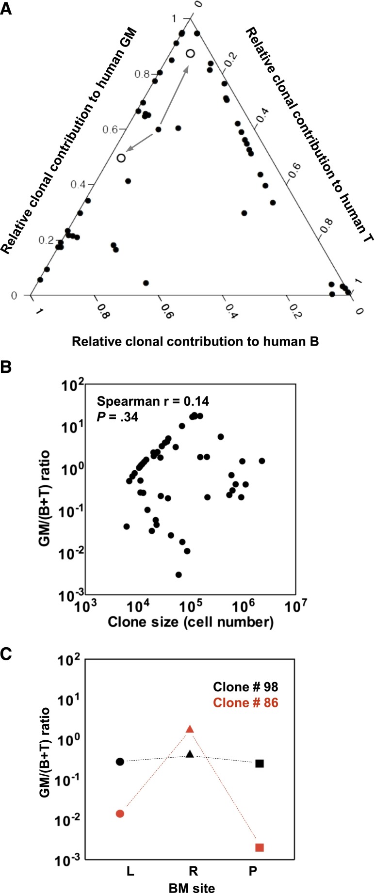
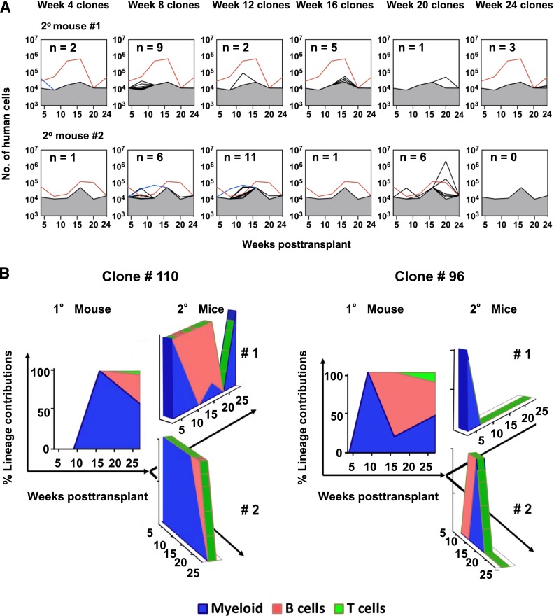
Human cord blood (CB) offers an attractive source of cells for clinical transplants because of its rich content of cells with sustained repopulating ability in spite of an apparent deficiency of cells with rapid reconstituting ability. Nevertheless, the clonal dynamics of nonlimiting CB transplants remain poorly understood. To begin to address this question, we exposed CD34+ CB cells to a library of barcoded lentiviruses and used massively parallel sequencing to quantify the clonal distributions of lymphoid and myeloid cells subsequently detected in sequential marrow aspirates obtained from 2 primary NOD/SCID-IL2Rγ(-/-) mice, each transplanted with ∼10(5) of these cells, and for another 6 months in 2 secondary recipients. Of the 196 clones identified, 68 were detected at 4 weeks posttransplant and were often lympho-myeloid. The rest were detected later, after variable periods up to 13 months posttransplant, but with generally increasing stability throughout time, and they included clones in which different lineages were detected. However, definitive evidence of individual cells capable of generating T-, B-, and myeloid cells, for over a year, and self-renewal of this potential was also obtained. These findings highlight the caveats and utility of this model to analyze human hematopoietic stem cell control in vivo.


Figures

Figure 1
Figure 1
Experimental design and analysis of clones detected. (A) Schematic outline of the MPG lentiviral vector that contained a 27-nucleotide non-coding DNA barcode sequence inserted downstream of the GFP reporter gene. The barcode sequence was designed with variable nucleotide doublets (NN) repeated 5 times, each separated by a constant nucleotide triplet sequence as previously described. (B) Schematic outline of the experimental design. CD34+ CB cells were transduced with the lentiviral barcode library for 6 hours in vitro, of which 105 cells (∼3.6 × 10⁴ barcoded cells) were then intravenously transplanted into each of 2 sublethally irradiated NSG mice. BM aspirates were collected, total human CD45+ analyzed, and myeloid, B-, and T-cell subsets of GFP+ and GFP- cells were also separately isolated by FACS. After 27 weeks, all BM cells were harvested separately from both legs and pelvis of both primary mice. From one mouse, half the cells were sorted for clonal analysis and the other half were transplanted intravenously into 2 secondary mice. From the second primary mouse, all harvested BM cells were sorted and used for clonal analysis. (C) Analysis of clonal dynamics in the 2 transplanted primary mice. The upper blue line denotes the total number of human hematopoietic cells in the BM of each mouse (assumed to have a total cellularity of 2 × 10⁸) and the upper green line denotes the total number of GFP+ cells in each mouse. Each color underneath denotes the total contribution of all of the lineages detected in each clone, as inferred from the BM sample analyzed. Black checked and shaded regions indicate the detection limit of the FACS (5 × 10⁴ cells) and MPS (variable) methods, respectively.
Figure 2
Figure 2
Kinetics of clone appearance, size, persistence, and lineage content. (A) Lines depict changes throughout time in the total size of each barcoded (GFP+) clone in primary mice, extrapolated from the BM sample and distinguishing those only seen at the time indicated in the panel (shown as red lines) vs those also detected at other times (shown as black lines). Gray shaded region denotes the limit of detection by MPS. Each pie chart shows the relative distributions of different types of clones present at different times (of the line plot, above) according to the diversity of lineages present at that time. (B) Different patterns of lineage content of clones present at 27 weeks posttransplant. 13 clonal patterns were defined based on the changes (absolute increases or decreases) detected in the lineage content of each clone through time as illustrated by the representative plots shown. Colored dotted lines denote the absence of specific cell lineages at the indicated detection limit.
Figure 3
Figure 3
Clonal contributions to the total human GFP+ GM, B- and T-cell compartments in all clones present at 27 weeks posttransplant. (A) Ternary plot showing the relative contribution to the GM, B-, and T-cell lineages of each clone present at 27 weeks posttransplant in the 2 primary mice (calculated as described in the text). Clone #110 persisted until weeks 20 and 24 posttransplant in the 2 secondary mice. Its contributions to the lineages assessed in both are indicated (arrows from clone #110 to the open circles, one for each of the secondary recipient mice). (B) Lack of correlation between GM/(B+T) value and clone size. (C) Examples of clones with similar and highly dissimilar GM/(B+T) ratio in different BM sites. The GM/(B+T) ratios of clone #98 (in black) and clone #86 (in red) in the BM of left leg (L, circles), right leg (R, triangles) and pelvis (P, squares) are shown.
Figure 4
Figure 4
Changes throughout time in the lineage content of clones regenerated in secondary mice. (A) Changes through time in the total size of each barcoded (GFP+) clone detected in the 2 secondary mice. Clone #110 (red line) and clone #96 (blue line) are shown separately. Gray shaded region denotes the limit of detection by MPS. (B) Relative contributions of GM, B-, and T-cells to clones #110 and #96 over time in the secondary mice as compared with the primary mouse in which they were first seen.

References

    1. Doulatov S, Notta F, Laurenti E, Dick JE. Hematopoiesis: a human perspective. Cell Stem Cell. 2012;10(2):120–136. - PubMed
    1. Bhatia M, Wang JC, Kapp U, Bonnet D, Dick JE. Purification of primitive human hematopoietic cells capable of repopulating immune-deficient mice. Proc Natl Acad Sci USA. 1997;94(10):5320–5325. - PMC - PubMed
    1. Conneally E, Cashman J, Petzer A, Eaves C. Expansion in vitro of transplantable human cord blood stem cells demonstrated using a quantitative assay of their lympho-myeloid repopulating activity in nonobese diabetic-scid/scid mice. Proc Natl Acad Sci USA. 1997;94(18):9836–9841. - PMC - PubMed
    1. Muller-Sieburg CE, Sieburg HB. Clonal diversity of the stem cell compartment. Curr Opin Hematol. 2006;13(4):243–248. - PubMed
    1. Jordan CT, Lemischka IR. Clonal and systemic analysis of long-term hematopoiesis in the mouse. Genes Dev. 1990;4(2):220–232. - PubMed

Publication types

MeSH terms