. 2013 Oct 9;54(10):6605-11.
doi: 10.1167/iovs.13-12196.

Delta-opioid receptors attenuate TNF-α-induced MMP-2 secretion from human ONH astrocytes

Affiliations

Delta-opioid receptors attenuate TNF-α-induced MMP-2 secretion from human ONH astrocytes

Naseem Akhter et al. Invest Ophthalmol Vis Sci. .

Abstract

Purpose: We examined the signaling mechanisms involved in δ-opioid-receptor agonist, SNC-121-mediated attenuation of TNF-α-induced matrix metalloproteinase-2 (MMP-2) secretion from human optic nerve head (ONH) astrocytes.

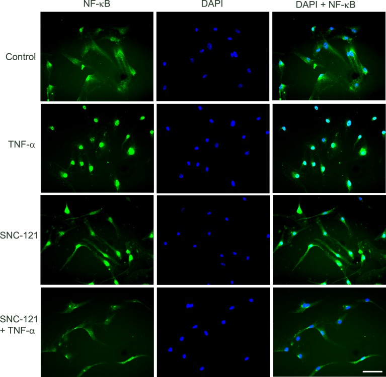
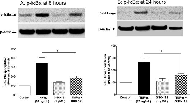
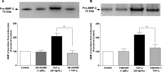
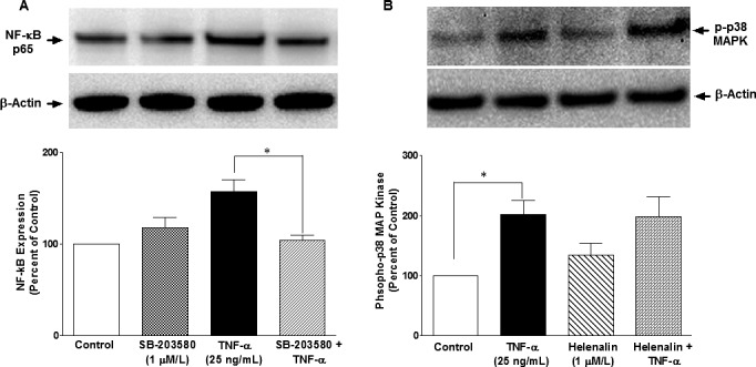
Methods: Human ONH astrocytes were treated with SNC-121 (1 μmol/L) for 15 minutes followed by TNF-α (25 ng/mL) treatment for 6 or 24 hours. Cells were pretreated with inhibitors of p38 mitogen-activated protein (MAP) kinase (SB-203580) or NF-κB (Helenalin) prior to TNF-α treatment. Changes in phosphorylation and expression of p38 MAP kinase, IκBα, NF-κB, and MMP-2 were measured by Western blotting. Translocation of NF-κB was determined by immunocytochemistry.

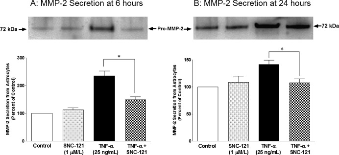
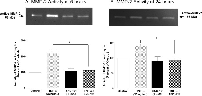
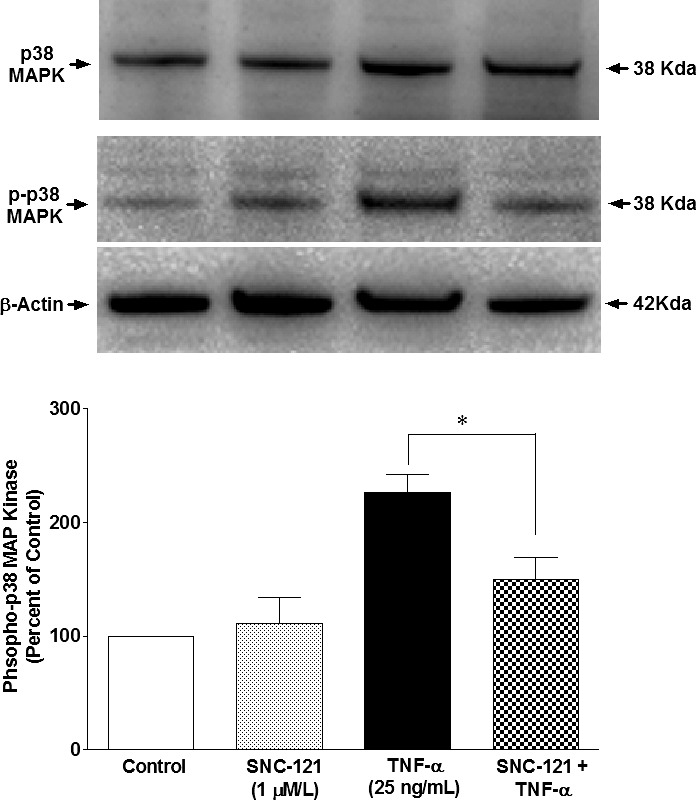
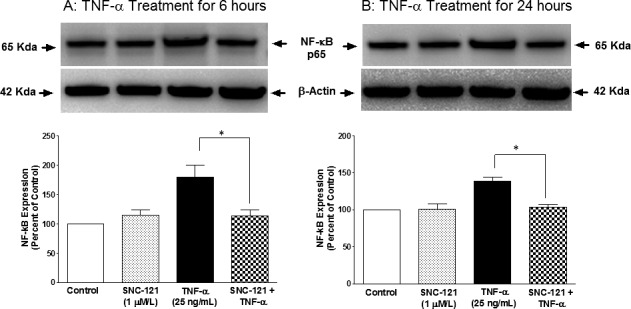
Results: TNF-α treatment increased MMP-2 secretion from ONH astrocytes to 236% ± 17% and 142% ± 8% at 6 and 24 hours, respectively; while SNC-121 treatment reduced MMP-2 secretion to 149% ± 11% and 108% ± 7% at 6 and 24 hours, respectively. The SNC-121-mediated inhibitory response was blocked by the δ-opioid-receptor antagonist naltrindole. TNF-α treatment resulted in a sustained phosphorylation of p38 MAP kinase up to 24 hours (226% ± 15% over control levels), which was reduced to 150% ± 20% by SNC-121 treatment. TNF-α treatment increased the expression of NF-κB to 179% ± 21% and 139% ± 6% at 6 and 24 hours, respectively, which was significantly blocked by SNC-121 treatment. Furthermore, TNF-α-induced MMP-2 secretion was blocked by 100% and 78% in the presence of SB-203580 and Helenalin, respectively.

Conclusions: Evidence is provided that SNC-121 attenuated TNF-α-induced MMP-2 secretion from ONH astrocytes. Data also supported the idea that p38 MAP kinase and NF-κB played central roles in TNF-α-induced MMP-2 secretion, and both were negatively regulated by SNC-121.

Keywords: astrocytes; glaucoma; matrix metalloproteinases; opioids; optic nerve head.


Figures

Figure 1
Figure 1
TNF-α–induced secretion of MMP-2 from ONH astrocytes at 6 (A) and 24 (B) hours. ONH astrocytes were starved overnight in serum-free medium. Cells were then pretreated with SNC-121 (1 μmol/L) for 15 minutes followed by TNF-α (25 ng/mL) treatment for 6 or 24 hours, respectively. Media was collected, concentrated 10-fold, and analyzed by Western blotting using selective anti-MMP-2 antibodies followed by incubation with appropriate secondary antibodies (HRP-conjugated; dilution 1:3000). The signal was captured using enhanced chemiluminescent reagent and a commercial imaging system (Bio-Rad). Data are expressed as mean ± SE. *P < 0.05. n = 6 to 7.
Figure 2
Figure 2
TNF-α–induced changes in MMP-2 activity in ONH astrocytes at 6 (A) and 24 (B) hours. ONH astrocytes were starved overnight in serum-free medium. Cells were then pretreated with SNC-121 (1 μmol/L) for 15 minutes followed by TNF-α (25 ng/mL) treatment for 6 or 24 hours, respectively. Media was collected, concentrated 10-fold, and analyzed by zymography as described in the Methods section. Data are expressed as mean ± SE. *P < 0.05. n = 4.
Figure 3
Figure 3
TNF-α–induced activation of p38 MAPK in ONH astrocytes. ONH astrocytes were starved overnight in serum-free medium. Cells were then pretreated with SNC-121 (1 μmol/L) for 15 minutes followed by TNF-α (25 ng/mL) treatment for 24 hours. Cell lysate (15 μg protein) was analyzed by Western blotting using selective anti-p38 MAPK or anti-phospho–p38 MAPK antibodies followed by incubation with appropriate secondary antibodies (HRP-conjugated; dilution 1:3000). The signal was captured using enhanced chemiluminescent reagent and the commercial imaging system (Bio-Rad). Data are expressed as mean ± SE. *P < 0.05. n = 4.
Figure 4
Figure 4
TNF-α–induced upregulation of NF-κB in ONH astrocytes. ONH astrocytes were starved overnight in serum-free medium. Cells were then pretreated with SNC-121 (1 μmol/L) for 15 minutes followed by TNF-α (25 ng/mL) treatment for 6 (A) or 24 (B) hours. Cell lysate (15 μg protein) was analyzed by Western blotting using selective anti–NF-κB antibodies followed by incubation with appropriate secondary antibodies (HRP-conjugated; dilution 1:3000). The signal was captured using enhanced chemiluminescent reagent and a commercial imaging system (Bio-Rad). Data are expressed as mean ± SE. *P < 0.05. n = 4.
Figure 5
Figure 5
TNF-α–induced translocation of NF-κB in ONH astrocytes at 6 hours. ONH astrocytes were starved in serum-free medium overnight. Cells were then pretreated with SNC-121 (1 μmol/L) for 15 minutes followed by TNF-α (25 ng/mL) treatment for 6 hours. ONH astrocytes were fixed by 4% paraformaldehyde followed by immunostaining with anti–NF-κB antibodies. The signal was captured by using AlexaFluor 488 conjugated secondary antibodies. Green color indicated staining for NF-κB and nuclear staining by DAPI was indicated by blue. There was no positive staining when primary antibodies were omitted (not shown). Fluorescence microscopy; bar size is 20 microns. Data shown in this figure are a representation of at least four independent experiments.
Figure 6
Figure 6
TNF-α–induced phosphorylation of IκBα in ONH astrocytes at 6 (A) and 24 (B) hours. ONH astrocytes were starved in serum-free medium overnight. Cells were then pretreated with SNC-121 (1 μmol/L) for 15 minutes followed by TNF-α (25 ng/mL) treatment for 6 or 24 hours. Cell lysate (15-μg protein) was analyzed by Western blotting using selective anti–phospho-IκBα antibodies followed by incubation with appropriate secondary antibodies (HRP-conjugated; dilution 1:3000). The signal was captured using enhanced chemiluminescent reagent and a commercial imaging system (Bio-Rad). Data are expressed as mean ± SE. *P < 0.05. n = 5 to 6.
Figure 7
Figure 7
Effects of SB-203580 (A) and Helenalin (B) on TNF-α–induced MMP-2 secretion from ONH astrocytes. ONH astrocytes were starved overnight in serum-free medium. Cells were then pretreated with a p38 MAPK inhibitor, SB-203580 (1 μmol/L), or NF-κB inhibitor, Helenalin (1 μmol/L) for 15 minutes followed by TNF-α (25 ng/mL) treatment for 6 hours. Media was collected, concentrated 10-fold, and analyzed by Western blotting using selective anti-MMP-2 antibodies followed by incubation with appropriate secondary antibodies (HRP-conjugated; dilution 1:3000). The signal was captured using enhanced chemiluminescent reagent and a commercial imaging system (Bio-Rad). Data are expressed as mean ± SE. *P < 0.05. n = 7 to 8.
Figure 8
Figure 8
Effects of SB-203580 (A) and Helenalin (B) on TNF-α–induced upregulation of NF-κB and p38 MAPK activation, respectively, in ONH astrocytes. ONH astrocytes were starved overnight in serum-free medium. Cells were then pretreated with SB203580 (1 μmol/L) or Helenalin (1 μmol/L) for 15 minutes followed by TNF-α (25 ng/mL) treatment for 6 hours. Cell lysate (15 μg protein) was analyzed by Western blotting using selective anti–NF-κB or anti–phospho-p38 MAP kinase antibodies followed by incubation with appropriate secondary antibodies (HRP-conjugated; dilution 1:3000). The signal was captured using enhanced chemiluminescent reagent and a commercial imaging system (Bio-Rad). Data are expressed as mean ± SE. *P < 0.05. n = 7 to 8.

References

    1. Hernandez MR. The optic nerve head in glaucoma: role of astrocytes in tissue remodeling. Prog Retin Eye Res. 2000; 19: 297– 321 - PubMed
    1. Hernandez MR, Pena JD. The optic nerve head in glaucomatous optic neuropathy. Arch Ophthalmol. 1997; 115: 389– 395 - PubMed
    1. Morrison JC, Dorman-Pease ME, Dunkelberger GR, Quigley HA. Optic nerve head extracellular matrix in primary optic atrophy and experimental glaucoma. Arch Ophthalmol. 1990; 108: 1020– 1024 - PubMed
    1. Toft-Hansen H, Fuchtbauer L, Owens T. Inhibition of reactive astrocytosis in established experimental autoimmune encephalomyelitis favors infiltration by myeloid cells over T cells and enhances severity of disease. Glia. 2011; 59: 166– 176 - PubMed
    1. Woessner JF Jr. Matrix metalloproteinases and their inhibitors in connective tissue remodeling. FASEB J. 1991; 5: 2145– 2154 - PubMed

Publication types

MeSH terms