. 2013 Dec;43(12):3219-32.
doi: 10.1002/eji.201343469. Epub 2013 Oct 14.

Identification of novel markers for mouse CD4(+) T follicular helper cells

Affiliations

Identification of novel markers for mouse CD4(+) T follicular helper cells

Smita S Iyer et al. Eur J Immunol. 2013 Dec.

Erratum in

  • Eur J Immunol. 2014 Jul;44(7):2197

Abstract

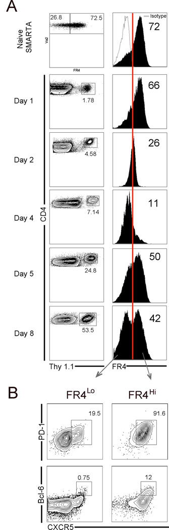
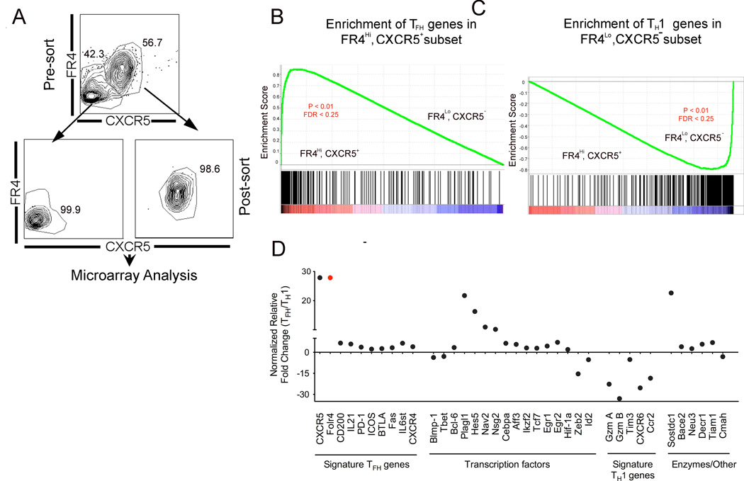
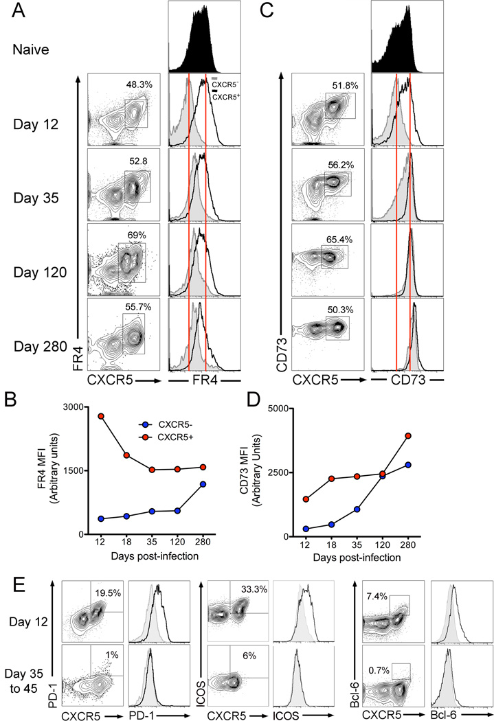
CD4(+) T follicular helper (TFH) cells are central for generation of long-term B-cell immunity. A defining phenotypic attribute of TFH cells is the expression of the chemokine R CXCR5, and TFH cells are typically identified by co-expression of CXCR5 together with other markers such as PD-1, ICOS, and Bcl-6. Herein, we report high-level expression of the nutrient transporter folate R 4 (FR4) on TFH cells in acute viral infection. Distinct from the expression profile of conventional TFH markers, FR4 was highly expressed by naive CD4(+) T cells, was downregulated after activation and subsequently re-expressed on TFH cells. Furthermore, FR4 expression was maintained, albeit at lower levels, on memory TFH cells. Comparative gene expression profiling of FR4(hi) versus FR4(lo) Ag-specific CD4(+) effector T cells revealed a molecular signature consistent with TFH and TH1 subsets, respectively. Interestingly, genes involved in the purine metabolic pathway, including the ecto-enzyme CD73, were enriched in TFH cells compared with TH1 cells, and phenotypic analysis confirmed expression of CD73 on TFH cells. As there is now considerable interest in developing vaccines that would induce optimal TFH cell responses, the identification of two novel cell surface markers should be useful in characterization and identification of TFH cells following vaccination and infection.

Keywords: B cells; CD4+ T cells; CD73; Folate R 4; Viral infection.


Conflict of interest statement

Conflicts of Interest

The authors have no conflicts of interest to disclose

Figures

Figure 1
Figure 1
FR4 expression distinguishes TFH and TH1 subsets. Antigen-specific CD4 effectors in spleen were sorted based on Ly6C expression and mRNA levels of FR4 (A) and other folate transport genes (B) were examined. (C) Flow cytometric analysis confirmed high FR4 expression on Ly6Clo antigen-specific CD4 T cells. (D) shows expression of CXCR5 and PD-1 on day 8 effectors and histograms show FR4 expression on TFH and TH1 subsets in. In (E) comparative phenotypic analysis of FR4hi, CXCR5+ (red histogram) and FR4lo, CXCR5 subsets (blue histogram) is shown. FR4 is highly expressed by GL7+, CXCR5+ GC TFH (F) and (G) shows localization of FR4 expressing CD4 T cells in the germinal centers in spleen at day 12 post-infection. (H) shows comparative expression profile of FR4 on antigen-specific CD4 T cells in the spleen, liver, and lung. FR4 expression on endogenous GP66 tetramer positive cells is shown in (I). Data are representative of three independent experiments.
Figure 2
Figure 2
FR4 expression on naive precursors does not determine TFH/TH1 commitment. Differentiation of FR4hi and FR4lo cells isolated from naive mouse spleen was determined as shown in (A). In (B), frequency and expression of FR4 on donor cells is shown in recipient spleen at day 8 post-infection. Bar graphs in (C) show number and frequency of donor cells per spleen. (D), shows % of TFH identified by co-expression of CXCR5 and PD-1, and CXCR5 and FR4 and % of GL7+ GC TFH in spleen at day 8 post-infection. Data are representative of three independent experiments. Bar graphs show mean ± SEM
Figure 3
Figure 3
Dynamic regulation of FR4 expression during TFH differentiation. (A) Kinetic analysis of FR4 expression on naive and antigen-specific CD4 T cells. Red line indicates percentage of FR4hi cells relative to isotype control staining. In (B), percentage of CXCR5+, PD-1hi TFH cells and CXCR5+, Bcl-6+ GC TFH cells in FR4lo versus FR4hi subsets is shown. Data are representative of three independent experiments.
Figure 4
Figure 4
Re-expression of FR4 on TFH cells requires interaction with cognate B cells. SMARTA CD4 T cells were transferred to WT or HEL Tg mice and responses were assessed 8 days after LCMV infection (A). Flow plots in (B) are gated on total B220+ B cells and show Fas+, PNA+, GC B cells in spleen. (C) shows % GC cells in spleen and total number of GC B cells in spleen is shown in (D). (E) LCMV enzyme linked immunosorbent assay (ELISA) was performed at day 8; LCMV-specific IgG endpoint titers in sera are shown. Flow plots in (F) are gated on CD4+ SMARTA cells in the spleen and show frequency of TFH cells and GC TFH cells in WT and HEL Tg mice. (G) shows frequency of FR4hi, CXCR5+ cells and black histogram shows % FR4hi antigen-specific CD4 T cells. Bar graph shows mean % TFH and % FR4hi antigen-specific CD4 T cells in peripheral blood (H) and spleen (I). (J), shows total number of TFH and FR4hi cells in spleen. Data are representative of two independent experiments. Bar graphs show mean ± SEM
Figure 5
Figure 5
Gene expression profiling of TFH and TH1 cells. (A) shows sorting strategy used to purify day 12 SMARTA CD4 T effectors based on FR4 and CXCR5 expression. Differentially regulated genes between TFH and TH1 subsets were identified using significance analysis of microarrays, which found 711 differentially regulated genes with a false discovery rate of 0.08 and a fold change of at least 2-fold. GSEA plots in B and C, show enrichment profile of TFH and TH1 genes in FR4hi, CXCR5hi and FR4lo, CXCR5lo subsets, respectively. Relative expression of select genes in TFH/TH1 subsets is shown in (D). Of note, CXCR5+, FR4hi cells did not express Foxp3 transcript levels indicating that this subset did not comprise of T follicular regulatory cells. Data are representative of two independent replicates.
Figure 6
Figure 6
Gene expression profiling leads to identification of CD73 as a novel TFH cell marker. Table A provides a list of select metabolic pathways enriched in TFH. (B), shows genes related to purine metabolic pathway differentially expressed between TFH and TH1 subsets; P2rx7, purinergic receptor P2X ligand gated ion channel 7; ADA, adenosine deaminase; ADK, adenosine kinase; Pde2a, phosphodiesterase 2a; Dck, deoxycytidine kinase. (C) Phenotypic analysis of CD73 confirms high expression on FR4hi and CXCR5+ antigen-specific TFH cells. (D, E) Comparison of TFH cells identified using CXCR5, PD-1 versus CD73 and FR4. Data are representative of three independent replicates.
Figure 7
Figure 7
Expression of FR4 is sustained on memory CXCR5+ antigen-specific cells. Kinetics of FR4 (A,B) and CD73 (C,D) expression on antigen-specific CD4 T cells from day 12 to day 280. Histograms show expression on CXCR5 (grey) and CXCR5+ (black) subsets. Figure E shows expression of PD-1, ICOS, and Bcl-6 on antigen-specific CD4 T cells at effector and memory time-points. Data are representative of three independent experiments

References

    1. Breitfeld D, Ohl L, Kremmer E, Ellwart J, Sallusto F, Lipp M, Forster R. Follicular B helper T cells express CXC chemokine receptor 5, localize to B cell follicles, and support immunoglobulin production. J Exp Med. 2000;192:1545–1552. - PMC - PubMed
    1. Berek C, Berger A, Apel M. Maturation of the immune response in germinal centers. Cell. 1991;67:1121–1129. - PubMed
    1. Linterman MA, Rigby RJ, Wong RK, Yu D, Brink R, Cannons JL, Schwartzberg PL, Cook MC, Walters GD, Vinuesa CG. Follicular helper T cells are required for systemic autoimmunity. J Exp Med. 2009;206:561–576. - PMC - PubMed
    1. Schaerli P, Willimann K, Lang AB, Lipp M, Loetscher P, Moser B. CXC chemokine receptor 5 expression defines follicular homing T cells with B cell helper function. J Exp Med. 2000;192:1553–1562. - PMC - PubMed
    1. Haynes NM, Allen CD, Lesley R, Ansel KM, Killeen N, Cyster JG. Role of CXCR5 and CCR7 in follicular Th cell positioning and appearance of a programmed cell death gene-1high germinal center-associated subpopulation. J Immunol. 2007;179:5099–5108. - PubMed

Publication types

MeSH terms

LinkOut - more resources