. 2013 Oct 15;128(16):1748-57.
doi: 10.1161/CIRCULATIONAHA.113.003313. Epub 2013 Sep 12.

Oxidized Ca(2+)/calmodulin-dependent protein kinase II triggers atrial fibrillation

Affiliations

Oxidized Ca(2+)/calmodulin-dependent protein kinase II triggers atrial fibrillation

Anil Purohit et al. Circulation. .

Abstract

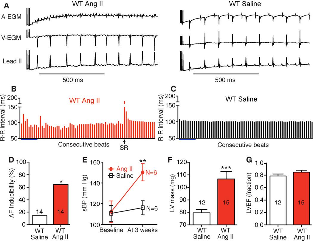
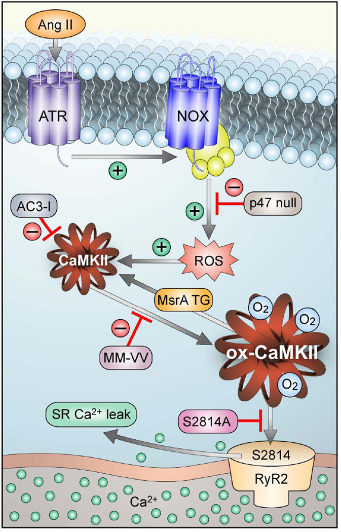
Background: Atrial fibrillation (AF) is a growing public health problem without adequate therapies. Angiotensin II and reactive oxygen species are validated risk factors for AF in patients, but the molecular pathways connecting reactive oxygen species and AF are unknown. The Ca(2+)/calmodulin-dependent protein kinase II (CaMKII) has recently emerged as a reactive oxygen species-activated proarrhythmic signal, so we hypothesized that oxidized CaMKIIδ could contribute to AF.

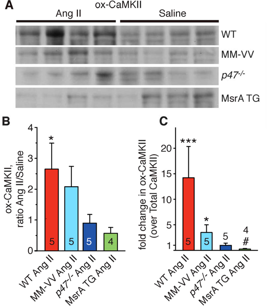
Methods and results: We found that oxidized CaMKII was increased in atria from AF patients compared with patients in sinus rhythm and from mice infused with angiotensin II compared with mice infused with saline. Angiotensin II-treated mice had increased susceptibility to AF compared with saline-treated wild-type mice, establishing angiotensin II as a risk factor for AF in mice. Knock-in mice lacking critical oxidation sites in CaMKIIδ (MM-VV) and mice with myocardium-restricted transgenic overexpression of methionine sulfoxide reductase A, an enzyme that reduces oxidized CaMKII, were resistant to AF induction after angiotensin II infusion.

Conclusions: Our studies suggest that CaMKII is a molecular signal that couples increased reactive oxygen species with AF and that therapeutic strategies to decrease oxidized CaMKII may prevent or reduce AF.

Keywords: angiotensin II; arrhythmias, cardiac; atrial fibrillation; calcium-calmodulin-dependent protein kinase type II; reactive oxygen species.


Conflict of interest statement

Conflict of Interest Disclosures: MEA is a cofounder of Allosteros Therapeutics, a biotech aiming to develop enzyme inhibitors to treat arrhythmias.

Figures

Figure 1
Figure 1
ox-CaMKII is increased in atria from patients with Atrial Fibrillation (AF). A. Representative immunofluorescence images using antiserum against ox-CaMKII in fixed sections of right atrial tissue from patients with sinus rhythm (SR) or AF. B. Image quantification showing significantly higher ox-CaMKII in patients with AF compared to SR (*p<0.05, Student’s t-test). C. Representative immunoblots with ox-CaMKII antiserum in right atrial tissue homogenates from patients in SR or AF. D. Quantification of immunoblots showing significantly higher ox-CaMKII expression in patients with AF compared to SR (*p<0.05, Student’s t-test). The % value indicates the mean ox-CaMKII/GAPDH ratio as normalized to the mean ox-CaMKII/GAPDH ratio in the SR group. E. Representative immunoblots with total CaMKII antiserum in right atrial tissue homogenates from patients in SR or AF. F. Quantification of immunoblots showing similar total CaMKII expression in patients with AF and SR (p=0.3, Student’s t-test). The % value indicates the mean CaMKII/GAPDH ratio as normalized to the mean CaMKII/GAPDH ratio in the SR group. The numerals shown in the bars indicate the sample size in each group, here and in subsequent figures.
Figure 2
Figure 2
Ang II treatment increases AF inducibility in WT mice. A. Representative atrial (AEGM) and ventricular (V-EGM) intracardiac electrograms and lead II surface ECG immediately after burst pacing show AF or SR in WT mice treated with Ang II or saline for 3 weeks. B. Contrasting R-R interval variability in AF and SR (C). Blue bars indicate calculated values from lead II ECGs shown in panel A. D. Higher AF inducibility in the Ang II treatment group (*p<0.05, Fisher’s exact test). E. Increase in systolic blood pressure (sBP) in WT mice after 3 weeks of Ang II treatment (**p<0.01, Student’s t-test). The numerals shown in the graph indicate the number of mice in each group. F. Significantly higher echocardiographically estimated left ventricular (LV) mass in Ang II treated mice compared to saline controls (***p<0.001, Student’s t-test). G. Similar LV ejection fraction (LVEF) in Ang II and saline treated mice.
Figure 3
Figure 3
CaMKII oxidation is critical to Ang II mediated AF. A. MM-VV, p47−/− and MsrA TG mice were resistant to Ang II mediated AF (*p<0.05 versus Ang II treated MM-VV, p47−/− and MsrA TG mice, Fisher’s exact test). B. All mice in panel A (WT, MM-VV, p47−/− and MsrA TG) showed a pressor response to Ang II. C. Ang II treatment induced cardiac hypertrophy as assessed by heart weight normalized to body weight (all comparisons versus saline controls from each genotype after 3 weeks of treatment; *p<0.05, ** p<0.01 and ***p<0.001, Student’s t-test).
Figure 4
Figure 4
ox-CaMKII in atria after Ang II or saline treatment. A. Atrial lysate immunoblots from WT, MM-VV, p47−/− and MsrA TG mice treated with Ang II or saline for 3 weeks and probed with an antiserum for ox-CaMKII. For quantification, ox-CaMKII bands were normalized to the total protein loading as assessed with Coomassie staining of the membrane. B. Increase in ox- CaMKII with Ang II treatment expressed as relative to the saline treated group. From each genotype 4 saline treated mice were used as controls. *p<0.05, for WT Ang II versus WT saline (*), in all other genotypes Ang II versus saline p>0.05; in addition, p=0.02 for WT Ang II versus MsrA TG Ang II and p=0.05 for MM-VV Ang II versus MsrA TG Ang II. C. Fold change in ox- CaMKII (over total CaMKII) in Ang II as relative to saline treated mice of the same genotype. From each genotype 4 saline treated mice were used as controls. ***p<0.001 versus WT saline, *p<0.05 versus MM-VV saline, #p<0.05 versus MsrA TG saline. WT Ang II versus p47−/− Ang II, P = 0.001, WT Ang II versus MsrA TG Ang II, P<.0001, MM-VV Ang II versus MsrA TG Ang II, P=0.001. Data were analyzed using two-way ANOVA (for treatment and genotype) with Bonferroni post-hoc comparisons.
Figure 5
Figure 5
Ang II promotes Ca2+ sparks and DADs. A. Representative examples of Ca2+ sparks in atrial myocytes from Ang II and saline treated WT and MM-VV mice. B. Summary of Ca2+ spark frequency data in atrial myocytes from Ang II treated mice compared to saline treated mice (*p<0.05 versus saline; Student’s t-test); WT saline (N=23 cells from 5 mice), WT Ang II (N=30 cells from 4 mice), MM-VV saline (N=36 cells from 4 mice) and MM-VV Ang II (N=28 cells from 4 mice). C. Examples of stimulated action potentials and a spontaneous, DAD triggered action potential. D. Higher incidence of DADs in atrial myocytes from Ang II treated WT mice (*p<0.05 versus saline, Fisher’s exact test) but not in Ang II treated MM-VV mice compared to saline controls. Numerals show cells with DADs/total cells studied for each group.
Figure 6
Figure 6
CaMKII activation and RyR2 serine 2814 are required for AF in Ang II infused mice. A. AC3-I and S2814A mice were treated with Ang II for 3 weeks and then burst paced to induce AF. AC3-I and S2814A mice were resistant to Ang II mediated AF promotion compared to WT Ang II treated mice (*p<0.05 versus all, Fisher’s Exact test, N=number of mice tested in each group). B. AC3-I and S2814A mice show similar systolic blood pressure (sBP) elevation after treatment with Ang II. Final sBP measurements were performed on three consecutive days prior to AF induction as shown in panel A. The numerals in the graph indicate the number of mice in each group. C. Ang II treatment causes similar cardiac hypertrophy in AC3-I and S2814A mice compared to saline controls (***p<0.001 versus AC3-I saline and **p=0.01 versus S2814A saline).
Figure 7
Figure 7
Schematic to illustrate the proposed mechanism of AF in Ang II infused mice. Ang II binding activates NADPH oxidase (NOX) to increase reactive oxygen species (ROS), leading to oxidation of methionines 281/282 in CaMKII (ox-CaMKII). Elevated ox-CaMKII phosphorylates serine 2814 on RyR2, causing enhanced diastolic Ca2+ leak that promotes AF triggering DADs. Genetically modified mice were used to test key steps of the proposed pathway.

Comment in

References

    1. Benjamin EJ, Wolf PA, D'Agostino RB, Silbershatz H, Kannel WB, Levy D. Impact of atrial fibrillation on the risk of death: the Framingham Heart Study. Circulation. 1998;98:946–952. - PubMed
    1. Khatib R, Joseph P, Briel M, Yusuf S, Healey J. Blockade of the renin-angiotensin-aldosterone system (RAAS) for primary prevention of non-valvular atrial fibrillation: A systematic review and meta analysis of randomized controlled trials. Int J Cardiol. 2013;165:17–24. - PubMed
    1. Nattel S. New ideas about atrial fibrillation 50 years on. Nature. 2002;415:219–226. - PubMed
    1. Shimano M, Shibata R, Inden Y, Yoshida N, Uchikawa T, Tsuji Y, Murohara T. Reactive oxidative metabolites are associated with atrial conduction disturbance in patients with atrial fibrillation. Heart Rhythm. 2009;6:935–940. - PubMed
    1. Neuman RB, Bloom HL, Shukrullah I, Darrow LA, Kleinbaum D, Jones DP, Dudley SC. Oxidative stress markers are associated with persistent atrial fibrillation. Clin Chem. 2007;53:1652–1657. - PMC - PubMed

Publication types

MeSH terms

Substances