

Microvascular abnormality in schizophrenia as shown by retinal imaging
- PMID: 24030514
- PMCID: PMC3857729
- DOI: 10.1176/appi.ajp.2013.13020234
Microvascular abnormality in schizophrenia as shown by retinal imaging
Abstract
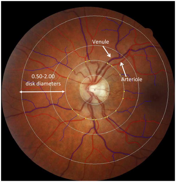
Objective: Retinal and cerebral microvessels are structurally and functionally homologous, but unlike cerebral microvessels, retinal microvessels can be noninvasively measured in vivo by retinal imaging. The authors tested the hypothesis that individuals with schizophrenia exhibit microvascular abnormality and evaluated the utility of retinal imaging as a tool for schizophrenia research.
Method: Participants were members of the Dunedin Study, a population-representative cohort followed from birth with 95% retention. Study members underwent retinal imaging at age 38. The authors assessed retinal arteriolar and venular caliber for all members of the cohort, including individuals who developed schizophrenia.
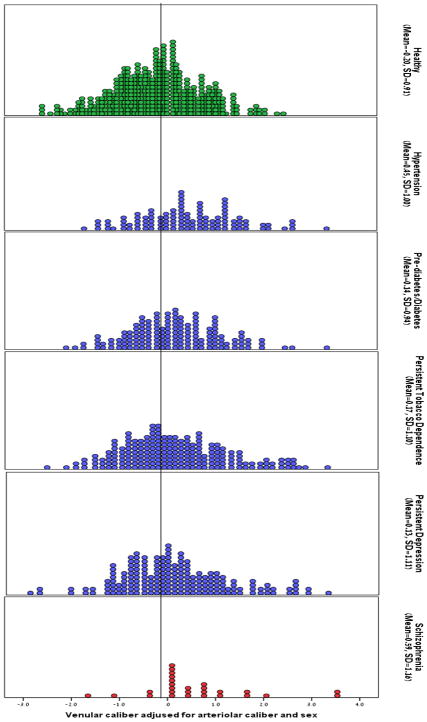
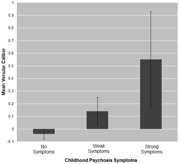
Results: Study members who developed schizophrenia were distinguished by wider retinal venules, suggesting microvascular abnormality reflective of insufficient brain oxygen supply. Analyses that controlled for confounding health conditions suggested that wider retinal venules are not simply an artifact of co-occurring health problems in schizophrenia patients. Wider venules were also associated with a dimensional measure of adult psychosis symptoms and with psychosis symptoms reported in childhood.
Conclusions: The findings provide initial support for the hypothesis that individuals with schizophrenia show microvascular abnormality. Moreover, the results suggest that the same vascular mechanisms underlie subthreshold symptoms and clinical disorder and that these associations may begin early in life. These findings highlight the promise of retinal imaging as a tool for understanding the pathogenesis of schizophrenia.
Conflict of interest statement
The authors have no conflicts of interest to report.
Figures
Comment in
-
Looking schizophrenia in the eye.Am J Psychiatry. 2013 Dec;170(12):1382-4. doi: 10.1176/appi.ajp.2013.13081136. Am J Psychiatry. 2013. PMID: 24306333 No abstract available.
References
-
- Meynert T. Psychiatrie: Klinik der erkrankungen des vorderhirns. Vienna, Braumüller: 1884.
-
- Sun C, Wang JJ, Mackey DA, Wong TY. Retinal vascular caliber: Systemic, environmental, and genetic associations. Surv Ophthalmol. 2009;54(1):74–95. - PubMed
-
- Cheung CYL, Ikram MK, Sabanayagam C, Wong TY. Retinal microvasculature as a model to study the manifestations of hypertension. Hypertension. 2012;60(5):1094. - PubMed
-
- Ikram MK, Ong YT, Cheung CY, Wong TY. Retinal vascular caliber measurements: Clinical significance, current knowledge and future perspectives. Ophthalmologica. 2012 Epub 2012/09/26. - PubMed
Publication types
MeSH terms
Grants and funding
LinkOut - more resources
Full Text Sources
Other Literature Sources
Medical
