. 2013 Sep 13;341(6151):1250-3.
doi: 10.1126/science.1240988.

Cytoplasmic LPS activates caspase-11: implications in TLR4-independent endotoxic shock

Affiliations

Cytoplasmic LPS activates caspase-11: implications in TLR4-independent endotoxic shock

Jon A Hagar et al. Science. .

Abstract

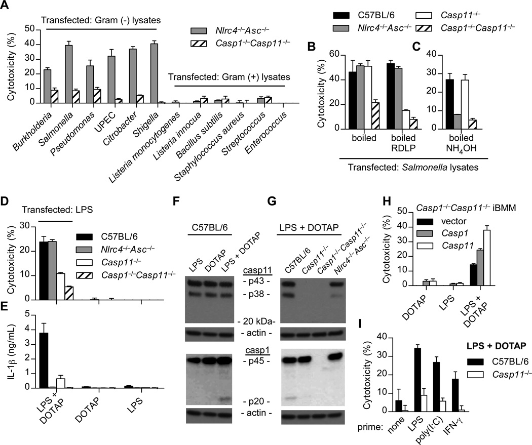
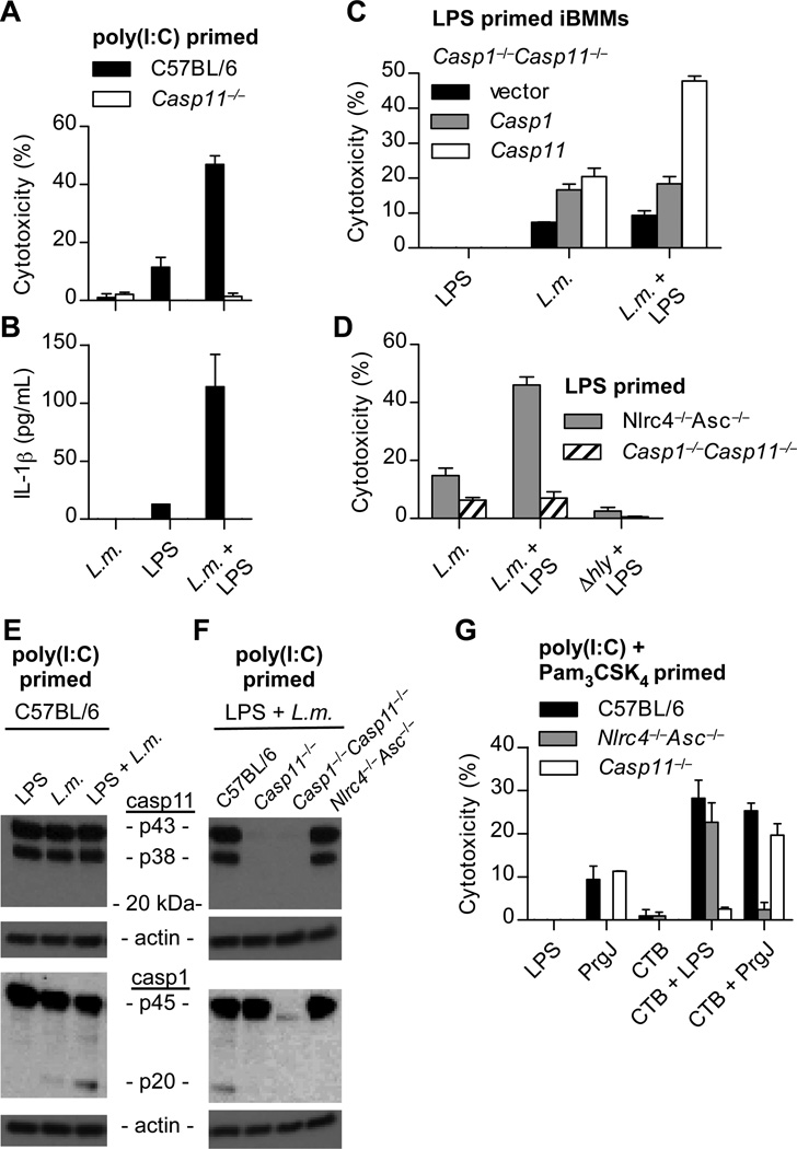
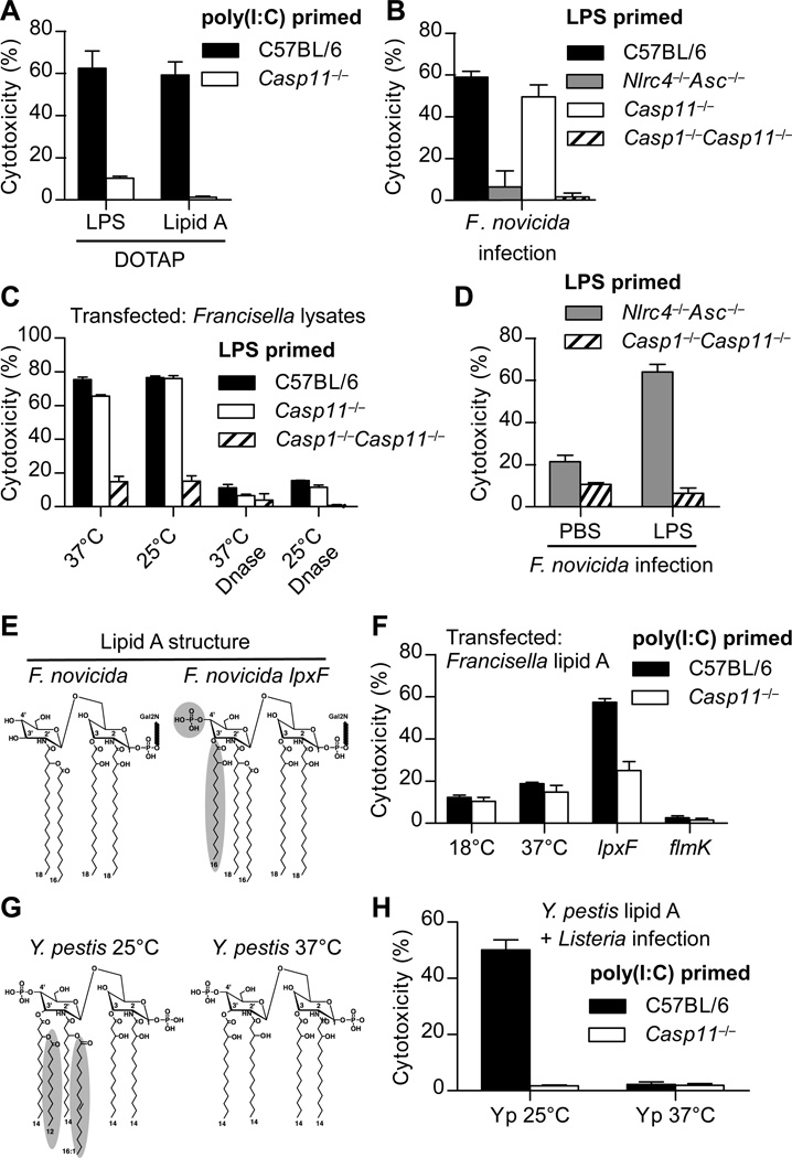
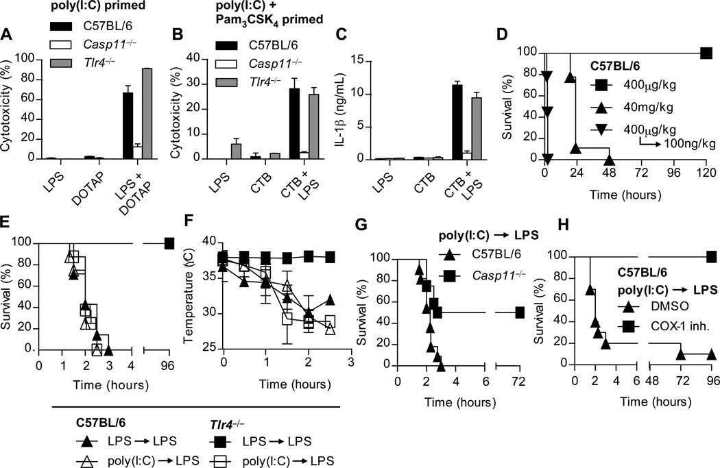
Inflammatory caspases, such as caspase-1 and -11, mediate innate immune detection of pathogens. Caspase-11 induces pyroptosis, a form of programmed cell death, and specifically defends against bacterial pathogens that invade the cytosol. During endotoxemia, however, excessive caspase-11 activation causes shock. We report that contamination of the cytoplasm by lipopolysaccharide (LPS) is the signal that triggers caspase-11 activation in mice. Specifically, caspase-11 responds to penta- and hexa-acylated lipid A, whereas tetra-acylated lipid A is not detected, providing a mechanism of evasion for cytosol-invasive Francisella. Priming the caspase-11 pathway in vivo resulted in extreme sensitivity to subsequent LPS challenge in both wild-type and Tlr4-deficient mice, whereas Casp11-deficient mice were relatively resistant. Together, our data reveal a new pathway for detecting cytoplasmic LPS.


Figures

Fig. 1
Fig. 1. Cytoplasmic LPS triggers caspase-11 activation
(A–H) BMM were LPS primed overnight prior to transfection. (A–C) BMMs were transfected with the indicated bacterial lysates packaged in Lipofectamine 2000. Cytotoxicity was determined by lactate dehydrogenase release 4 hours later. Where indicated, lysates were treated with RNase, DNase, proteinase K, and lysozyme (RDLP) (B) or ammonium hydroxide (C). (D–E) BMMs were transfected with ultrapure LPS from S. minnesota RE595 packaged with DOTAP, a liposomal transfection reagent. Cytotoxicity (D) and IL-1β secretion by ELISA (E) were determined 4 h post transfection. (F–G) BMMs were stimulated as in (D) and caspase-1 and -11 processing by western blot were examined 2 h post transfection. (H) Immortalized Casp1−/−Casp11−/− BMMs (iBMMs) complemented by retroviral transduction of Casp1 or Casp11 were transfected with LPS from S. minnesota RE595. Cytotoxicity was determined after 4 h. (I) Macrophages were primed overnight with LPS (50ng/mL), poly(I:C) (1µg/mL), IFN-γ (8ng/mL), or left untreated. Cells were then transfected with LPS from S. minnesota RE595 and cytotoxicity was determined 2 h later. Data are representative of at least 3 experiments. Error bars indicate standard deviation of technical replicates.
Fig. 2
Fig. 2. Listeria and CTB mediate caspase-11 activation by LPS
(A–F) The indicated macrophages were primed with poly(I:C) or LPS and then infected by L. monocytogenes (MOI 5) in the presence or absence of LPS from S. minnesota RE595 (1µg/mL). Cytotoxicity (A, C, D), IL-1β secretion (B), or caspase-1 and caspase-11 processing (E–F) were examined 4 h post-infection. (G) Poly(I:C) and Pam3CSK4 primed macrophages were incubated with the indicated combinations of CTB (20µg/mL), LPS from E. coli O111:B4 (1µg/mL), and PrgJ (10µg/mL). Cytotoxicity was determined 16 h later. Data are representative of 3 (A, D, G) or 2 (B, C, E, F) experiments. Error bars indicate standard deviation of technical replicates.
Fig. 3
Fig. 3. Caspase-11 responds to distinct lipid A structures
(A) Poly(I:C) primed BMMs were transfected with LPS from S. minnesota RE595 or S. typhimurium lipid A. Cytotoxicity was determined after 2 h. (B) Cytotoxicity in LPS primed BMMs was determined 4 hours after infection with F. novicida (MOI 200). (C) LPS primed BMMs were transfected with mock or DNase treated F. novicida lysates. Cytotoxicity was determined 4 hours later. (D) Macrophages were infected as in (B) in the presence or absence of LPS from S. minnesota RE595. (E) Structural comparison of lipid A from wild type F. novicida or the lpxF mutant. Structural changes are indicated. (F) Poly(I:C) primed macrophages were transfected with lipid A from F. novicida grown at 18°C or 37°C, or the indicated F. novicida mutants grown at 37°C. Cytotoxicity was determined after 2 h. (G) Structural comparison of lipid A from Y. pestis grown at 25°C or 37°C. (H) Poly(I:C) primed BMMs were infected with L. monocytogenes in the presence of lipid A from Y. pestis grown at 25°C or 37°C. Cytotoxicity was determined after 4 h. Data are representative of at least 3 (A, F, G) or 2 (B, C, D) experiments. Error bars indicate standard deviation of technical replicates.
Fig. 4
Fig. 4. LPS detection and rapid induction of shock in primed mice occur independently of TLR4
(A) BMMs were primed overnight with poly(I:C) and then transfected with S. minnesota RE595 LPS. Cytotoxicity was determined 2 hours later. (B–C) Poly(I:C) and Pam3CSK4 primed macrophages were incubated with the indicated combinations of CTB (20µg/mL) and LPS from E. coli O111:B4 (1µg/mL). Cytotoxicity (B) and IL-1β secretion (C) were determined 16 hours later. Data are representative of at least 3 experiments; error bars indicate standard deviation of technical replicates (A–C). (D) Survival of mice challenged with the indicated doses of Escherichia coli LPS, or primed with LPS and then re-challenged 7 hours later. Data are pooled from three experiments; n = 9 per condition. (E) Survival of mice primed with LPS (400μg/kg) or poly(I:C) (10µg/kg) and then challenged 7 hours later with LPS (100ng/kg). Data are pooled from three experiments; n = 7 per LPS prime group and n = 8 for poly(I:C) prime group. (F) Rectal temperatures of mice in panel (E) after LPS challenge. Data are representative of 3 experiments; n = 4 per condition. (G) Survival of poly(I:C) primed mice challenged 6 hours later with LPS (10ng/kg). Data are pooled from 3 experiments, n = 11 (C57BL/6) or 12 (Casp11−/−). (H) Mice were primed with poly(I:C) and then challenged 6 hours later with LPS (100ng/kg) and monitored for survival. 1 h before LPS challenge, mice were given 5mg/kg of COX-1 inhibitor or DMSO control. Data are pooled from 2 experiments; n = 11 per condition.

Comment in

  • Immunology. Sensing endotoxins from within.
    Kagan JC. Kagan JC. Science. 2013 Sep 13;341(6151):1184-5. doi: 10.1126/science.1243939. Science. 2013. PMID: 24031006 Free PMC article. No abstract available.
  • When LPS sneaks into the cell.
    Papatriantafyllou M. Papatriantafyllou M. Nat Rev Immunol. 2013 Nov;13(11):772-3. doi: 10.1038/nri3546. Epub 2013 Sep 30. Nat Rev Immunol. 2013. PMID: 24077101 No abstract available.

References

    1. Von Moltke J, Ayres JS, Kofoed EM, Chavarría-Smith J, Vance RE. Recognition of bacteria by inflammasomes. Annu. Rev. Immunol. 2013;31:73–106. - PubMed
    1. Masters SL, et al. NLRP1 Inflammasome Activation Induces Pyroptosis of Hematopoietic Progenitor Cells. Immunity. 2012;37:1009–1023. - PMC - PubMed
    1. Kayagaki N, et al. Non-canonical inflammasome activation targets caspase-11. Nature. 2011;479:117–121. - PubMed
    1. Aachoui Y, et al. Caspase-11 Protects Against Bacteria That Escape the Vacuole. Science. 2013;339:975–978. - PMC - PubMed
    1. Broz P, et al. Caspase-11 increases susceptibility to Salmonella infection in the absence of caspase-1. Nature. 2012;490:288–291. - PMC - PubMed

Publication types