Tubular-trabecular type Basal cell adenoma of the parotid gland: a patient report
- PMID: 24031120
- PMCID: PMC3763784
Tubular-trabecular type Basal cell adenoma of the parotid gland: a patient report
Abstract
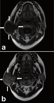
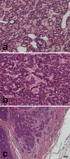
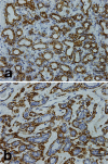
Basal cell adenoma (BCA) is an uncommon benign salivary gland neoplasm that includes isomorphic basaloid cells. We report on a female patient with BCA that developed in the right parotid gland in her 50s. The present patient demonstrated a few tumor nests in the fibrous capsule, and her tumor was larger than usual. These facts made us suspect of malignancy. Histopathologically, the tumor was characterized by multiple duct-like structures and tubular-trabecular masses composed of small isomorphic cells with hyperchromatic, round nuclei and an eosinophilic cytoplasm. It was difficult to determine whether the ductal structures noted in the tumor capsule were invasive. By immunohistochemistry, tumor cells of the tubular nests were positive for cytokeratin 7 and that the outer cells of tubular nests were positive for alpha smooth muscle actin (αSMA) and calponin. Tumor cells were immuno-negative for S-100 protein and glial fibrillary acidic protein. The Ki-67 labeling scores of the cells were extremely low (< 1%). We could achieve an accurate diagnosis of BCA by immunohistochemistry with MIB-1 and other markers.
Keywords: basal cell adenoma; immunohistochemistry; parotid gland.
Figures


References
-
- Barnes L, Eveson JW, Reichart P, Sidransky D. World Health Organization classification of tumours. Pathology and genetics of head and neck tumours Lyon : International Agency for Research on Cancer Press; 2005. p. 261–262
-
- Dardick I, Daley TD, van Nostrand AW. Basal cell adenoma with myoepithelial cell-derived "stroma": a new major salivary gland tumor entity. Head Neck Surg 1986; 8: 257–267 - PubMed
-
- Ellis GL, Auclair PL.Tumor of the salivary glands. Atlas of tumor pathology. Washington DC: Armed Forces Institute of Pathology; 1996. p. 90 –94
-
- Evenson JW, Cawson RA. Tumours of the minor (oropharyngeal) salivary glands: a demographic study of 336 cases. J Oral Pathol 1985; 14: 500–509 - PubMed
-
- Gnepp DR. Diagnostic surgical pathology of the head and neck. London: W. B. Saunders; 2009. p. 351–365
LinkOut - more resources
Full Text Sources
Research Materials
