. 2013 Sep 2:7:496.
doi: 10.3389/fnhum.2013.00496. eCollection 2013.

Applying independent component analysis to clinical FMRI at 7 t

Affiliations

Applying independent component analysis to clinical FMRI at 7 t

Simon Daniel Robinson et al. Front Hum Neurosci. .

Abstract

Increased BOLD sensitivity at 7 T offers the possibility to increase the reliability of fMRI, but ultra-high field is also associated with an increase in artifacts related to head motion, Nyquist ghosting, and parallel imaging reconstruction errors. In this study, the ability of independent component analysis (ICA) to separate activation from these artifacts was assessed in a 7 T study of neurological patients performing chin and hand motor tasks. ICA was able to isolate primary motor activation with negligible contamination by motion effects. The results of General Linear Model (GLM) analysis of these data were, in contrast, heavily contaminated by motion. Secondary motor areas, basal ganglia, and thalamus involvement were apparent in ICA results, but there was low capability to isolate activation in the same brain regions in the GLM analysis, indicating that ICA was more sensitive as well as more specific. A method was developed to simplify the assessment of the large number of independent components. Task-related activation components could be automatically identified via these intuitive and effective features. These findings demonstrate that ICA is a practical and sensitive analysis approach in high field fMRI studies, particularly where motion is evoked. Promising applications of ICA in clinical fMRI include presurgical planning and the study of pathologies affecting subcortical brain areas.

Keywords: artifacts; independent component analysis; motion; motor; neurology; presurgical planning; ultra-high field fMRI.


Figures

Figure 1
Figure 1
A schematic representation of data processing steps for each patient in the main analysis.
Figure 2
Figure 2
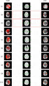
A comparison of GLM and ICA analyses of 7 T fMRI data with a chin task. GLM results are contaminated by motion artifacts (yellow and cyan arrows). ICA components show no motion contamination and bilateral activation throughout primary motor areas. Activated areas not present in corresponding GLM results, or not distinguishable from artifacts, are indicated by magenta arrows. White vertical lines separate sample slices covering the basal ganglia from those showing primary motor regions. All brain images are displayed in radiological convention.
Figure 3
Figure 3
Examination of the activation visible in GLM results over a range of thresholds (chin task). Activation visible in independent components and GLM results is compared in a single slice. The threshold corresponding to a GLM cluster-corrected P = 0.05 is indicated by a yellow spot. Activation maps are illustrated at higher and lower thresholds than this to allow the ability to separate activation and motion in GLM results to be assessed. Clusters which are substantially better defined in ICA are indicated by green arrows.
Figure 4
Figure 4
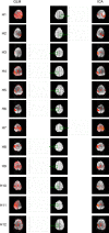
A comparison of GLM and ICA analyses of 7 T fMRI data with a hand task. The same thresholds were applied as in Figure 2. Activation in the basal ganglia and thalamus is indicated by arrows in ICA. Activated areas not present in corresponding GLM results, or not distinguishable from artifacts, are indicated by magenta arrows.
Figure 5
Figure 5
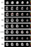
Additional motor components identified in the ICA results of chin patients C1, C2, C3, C6, C9, C10, H4, and H9.
Figure 6
Figure 6
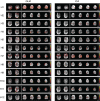
Mean responses in voxels in primary motor areas in the chin task. Time courses in PMA in C2 and C3 (outlined in red) are non-model-conform, and correspond with low sensitivity in GLM results (left) but not ICA (right).
Figure 7
Figure 7
Mean responses in voxels in primary motor areas in the hand task. Time courses in PMA in C2 and C3 (outlined in red) are non-model-conform, and correspond with low sensitivity in GLM results (left) but not ICA (right).
Figure 8
Figure 8
Motion artifact components isolated in data from Chin patient C5. These artefacts are attributed to motion in the anterior-posterior direction (A, B), through-plane motion (C), fluctuating Nyquist ghost (D, E).
Figure 9
Figure 9
A plot of the two most successful features used to automatically identify primary motor activation components (red circles) amongst other components (black crosses) in the Chin and Hand groups.

Similar articles

Cited by

References

    1. Abou-Elseoud A., Starck T., Remes J., Nikkinen J., Tervonen O., Kiviniemi V. (2010). The effect of model order selection in group PICA. Hum. Brain Mapp. 31, 1207–121610.1002/hbm.20929 - DOI - PMC - PubMed
    1. Barth M., Poser B. A. (2011). Advances in high-field BOLD fMRI. Materials 4, 1941–195510.3390/ma4111941 - DOI - PMC - PubMed
    1. Beckmann C. F. (2012). Modelling with independent components. Neuroimage 62, 891–90110.1016/j.neuroimage.2012.02.020 - DOI - PubMed
    1. Beckmann C. F., Deluca M., Devlin J. T., Smith S. M. (2005). Investigations into resting-state connectivity using independent component analysis. Philos. Trans. R. Soc. Lond. B Biol. Sci. 360, 1001–101310.1098/rstb.2005.1634 - DOI - PMC - PubMed
    1. Beckmann C. F., Smith S. M. (2004). Probabilistic independent component analysis for functional magnetic resonance imaging. IEEE Trans. Med. Imaging 23, 137–15210.1109/TMI.2003.822821 - DOI - PubMed

LinkOut - more resources