

Sonographic localization of a nonpalpable shunt: Ultrasound-assisted ventricular shunt tap
- PMID: 24032076
- PMCID: PMC3766327
- DOI: 10.4103/2152-7806.116151
Sonographic localization of a nonpalpable shunt: Ultrasound-assisted ventricular shunt tap
Abstract
Background: Patients frequently present to the emergency department (ED) for evaluation of cerebrospinal fluid (CSF) shunt malfunction, often requiring urgent management. A typical evaluation in the emergency room setting includes a thorough history and physical examination, noncontrasted head computed tomography (CT), shunt series, and occasionally a ventricular shunt tap.
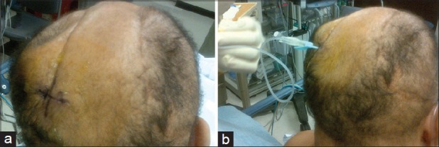
Case description: We present the case of a 53-year-old male who initially presented to the ED in acute status epilepticus. His history was notable for seizures and multiple craniectomies and cranioplasties with subsequent placement of a ventriculoperitoneal shunt secondary to traumatic brain injury. Imaging in the ED suggested possible shunt failure. No previous imaging was available for comparison, and therefore a ventricular shunt tap was attempted. Initially, the tap could not be performed, as the shunt was not palpable secondary to the thickness of his scalp and location of the reservoir near his complex cranial reconstruction site. We report, for the first time, the utility of emergency ultrasound (EUS) to aid in such an encounter.
Conclusion: EUS revealed the exact location of his shunt reservoir, and therefore enabled the shunt tap, which ultimately led to the discovery of the patient's proximal shunt failure in a setting that may have otherwise been missed. The patient underwent urgent shunt revision with a good outcome.
Keywords: Hydrocephalus; shunt failure; ultrasound guidance; ventricular shunt tap.
Figures


References
-
- Bondurant CP, Jimenez DF. Epidemiology of cerebrospinal fluid shunting. Pediatr Neurosurg. 1995;23:254–8. - PubMed
-
- Chan KH, Mann KS. Prolonged therapeutic external ventricular drainage. A prospective study. Neurosurgery. 1988;23:436–8. - PubMed
-
- de Oliveira RS, Machado HR. Transcranial color-coded Doppler ultrasonography for evaluation of children with hydrocephalus. Neurosurg Focus. 2003;15:ECP3. - PubMed
-
- Hamburg LM, Kessler DO. Rapid evaluation of ventriculoperitoneal shunt function in a pediatric patient using emergency ultrasound. Pediatr Emerg Care. 2012;28:726–7. - PubMed
-
- Iskandar BJ, McLaughlin C, Mapstone TB, Grabb PA, Oakes WJ. Pitfalls in the diagnosis of ventricular size. Pediatrics. 1998;101:1031–6. - PubMed
Publication types
LinkOut - more resources
Full Text Sources
Other Literature Sources
Miscellaneous
