

Non-invasive detection of neurochemical changes prior to overt pathology in a mouse model of spinocerebellar ataxia type 1
- PMID: 24032423
- PMCID: PMC4094372
- DOI: 10.1111/jnc.12435
Non-invasive detection of neurochemical changes prior to overt pathology in a mouse model of spinocerebellar ataxia type 1
Abstract
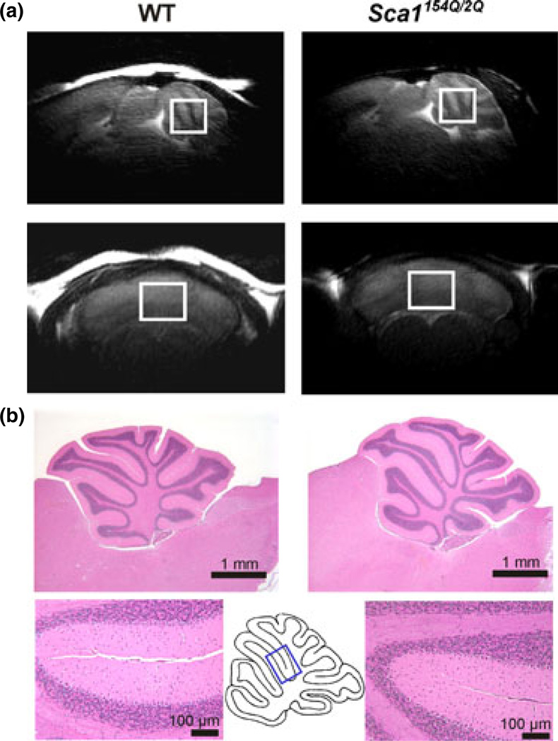
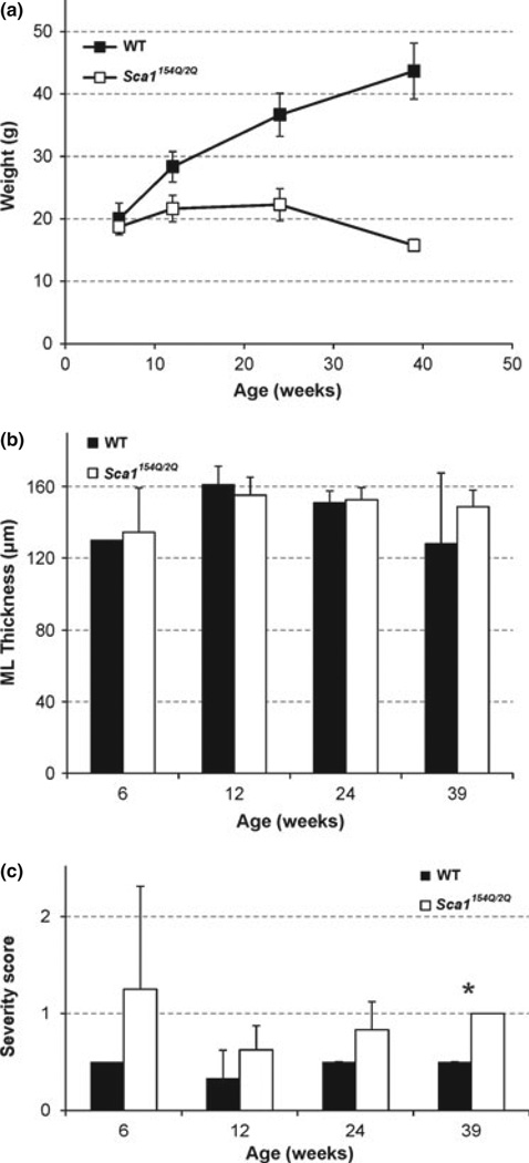
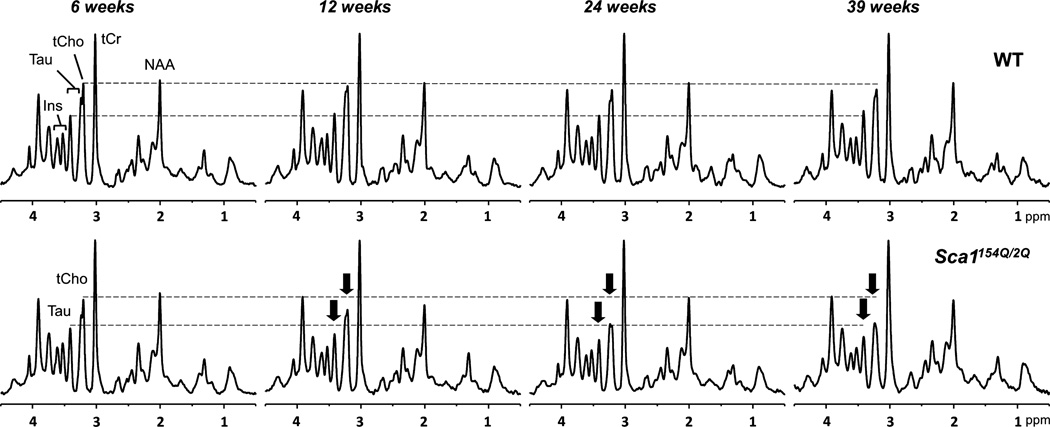
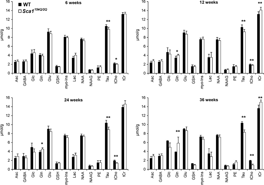
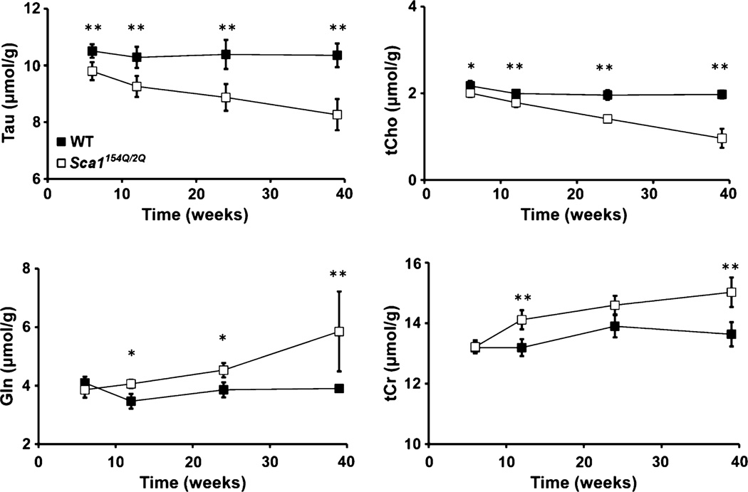
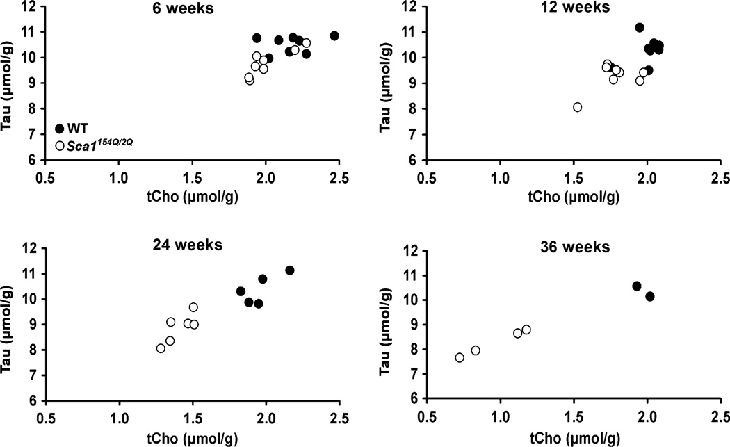
Spinocerebellar ataxia type 1 (SCA1) is a hereditary, progressive and fatal movement disorder that primarily affects the cerebellum. Non-invasive imaging markers to detect early disease in SCA1 will facilitate testing and implementation of potential therapies. We have previously demonstrated the sensitivity of neurochemical levels measured by (1) H magnetic resonance spectroscopy (MRS) to progressive neurodegeneration using a transgenic mouse model of SCA1. In order to investigate very early neurochemical changes related to neurodegeneration, here we utilized a knock-in mouse model, the Sca1(154Q/2Q) line, which displays milder cerebellar pathology than the transgenic model. We measured cerebellar neurochemical profiles of Sca1(154Q/2Q) mice and wild-type littermates using 9.4T MRS at ages 6, 12, 24, and 39 weeks and assessed the cerebellar pathology of a subset of the mice at each time point. The Sca1(154Q/2Q) mice displayed very mild cerebellar pathology even at 39 weeks, however, were distinguished from wild types by MRS starting at 6 weeks. Taurine and total choline levels were significantly lower at all ages and glutamine and total creatine levels were higher starting at 12 weeks in Sca1(154Q/2Q) mice than controls, demonstrating the sensitivity of neurochemical levels to neurodegeneration related changes in the absence of overt pathology. We measured cerebellar neurochemical alterations in a knock-in mouse model of spinocerebellar ataxia type 1, a hereditary movement disorder, using ultra-high field magnetic resonance spectroscopy (MRS). Very early neurochemical alterations were detectable prior to overt pathology in the volume-of-interest for MRS. Alterations were indicative of osmolytic changes and of disturbances in membrane phospholipid and energy metabolism.
Keywords: SCA1; cerebellum; histology; magnetic resonance spectroscopy; mouse model; neurodegeneration.
© 2013 International Society for Neurochemistry.
Conflict of interest statement
The authors declare no conflict of interest.
Figures
Comment in
-
Early impairment in brain metabolism detected by MR spectroscopy antedates structural changes in mouse models of spinocerebellar ataxias.J Neurochem. 2013 Dec;127(5):578-9. doi: 10.1111/jnc.12448. Epub 2013 Oct 23. J Neurochem. 2013. PMID: 24147560 No abstract available.
References
-
- Burright EN, Clark HB, Servadio A, Matilla T, Feddersen RM, Yunis WS, Duvick LA, Zoghbi HY, Orr HT. SCA1 transgenic mice: a model for neurodegeneration caused by an expanded CAG trinucleotide repeat. Cell. 1995;82:937–948. - PubMed
-
- Clark HB, Burright EN, Yunis WS, Larson S, Wilcox C, Hartman B, Matilla A, Zoghbi HY, Orr HT. Purkinje cell expression of a mutant allele of SCA1 in transgenic mice leads to disparate effects on motor behaviors, followed by a progressive cerebellar dysfunction and histological alterations. J. Neurosci. 1997;17:7385–7395. - PMC - PubMed
-
- Eder K, Kish SJ, Kirchgessner M, Ross BM. Brain phospholipids and fatty acids in Friedreich’s ataxia and spinocerebellar atrophy type-1. Mov. Disord. 1998;13:813–819. - PubMed
-
- Garwood M, DelaBarre L. The return of the frequency sweep: designing adiabatic pulses for contemporary NMR. J. Magn. Reson. 2001;153:155–177. - PubMed
Publication types
MeSH terms
Substances
Grants and funding
LinkOut - more resources
Full Text Sources
Other Literature Sources
