. 2013 Oct 9;135(40):15153-64.
doi: 10.1021/ja407059n. Epub 2013 Sep 26.

Monothiol glutaredoxins can bind linear [Fe3S4]+ and [Fe4S4]2+ clusters in addition to [Fe2S2]2+ clusters: spectroscopic characterization and functional implications

Affiliations

Monothiol glutaredoxins can bind linear [Fe3S4]+ and [Fe4S4]2+ clusters in addition to [Fe2S2]2+ clusters: spectroscopic characterization and functional implications

Bo Zhang et al. J Am Chem Soc. .

Abstract

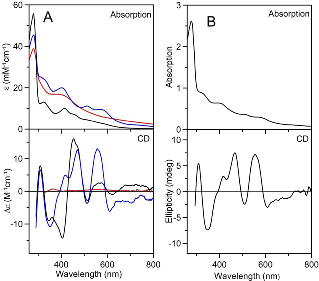
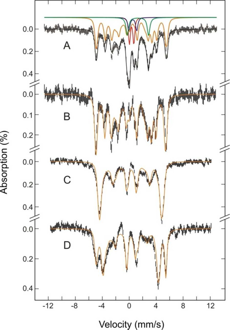
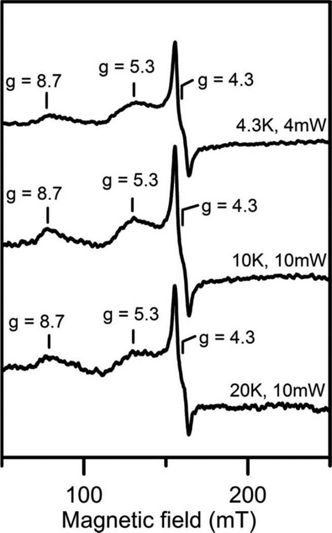
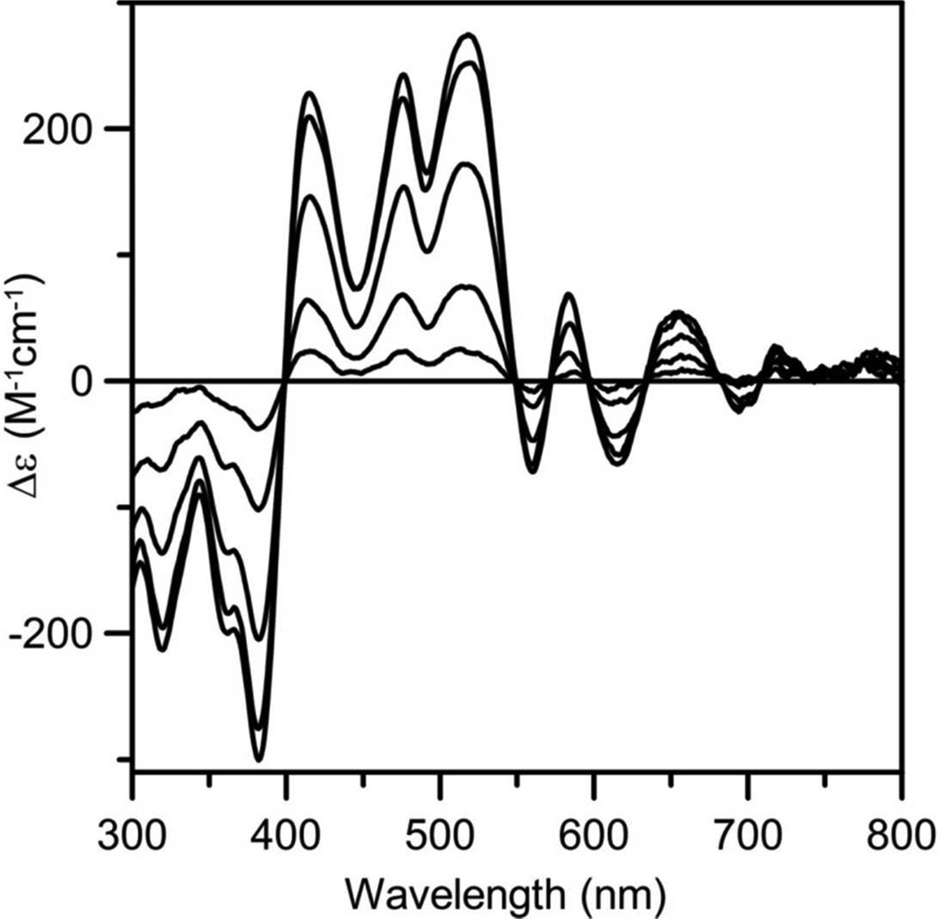
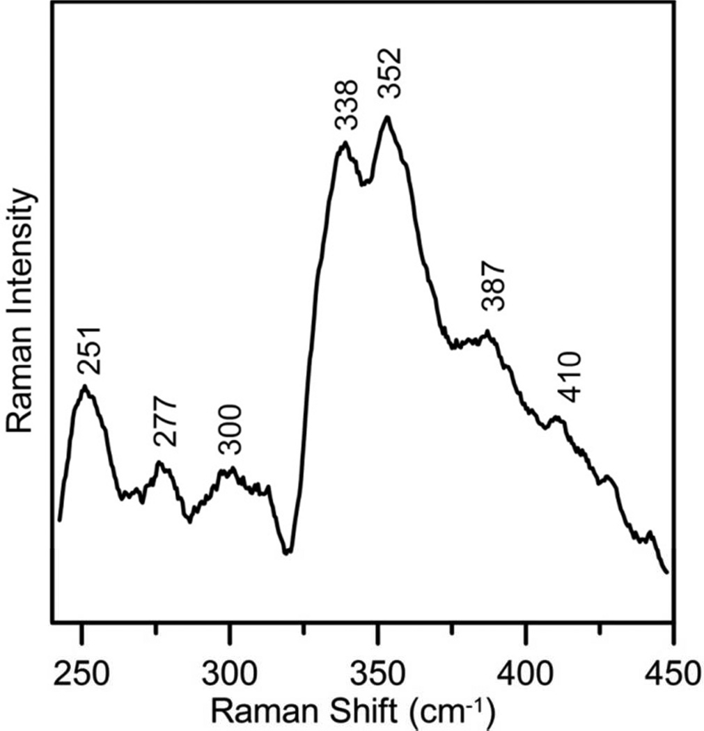
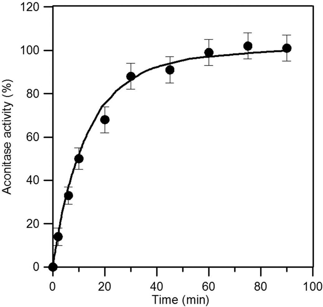
Saccharomyces cerevisiae mitochondrial glutaredoxin 5 (Grx5) is the archetypical member of a ubiquitous class of monothiol glutaredoxins with a strictly conserved CGFS active-site sequence that has been shown to function in biological [Fe2S2](2+) cluster trafficking. In this work, we show that recombinant S. cerevisiae Grx5 purified aerobically, after prolonged exposure of the cell-free extract to air or after anaerobic reconstitution in the presence of glutathione, predominantly contains a linear [Fe3S4](+) cluster. The excited-state electronic properties and ground-state electronic and vibrational properties of the linear [Fe3S4](+) cluster have been characterized using UV-vis absorption/CD/MCD, EPR, Mössbauer, and resonance Raman spectroscopies. The results reveal a rhombic S = 5/2 linear [Fe3S4](+) cluster with properties similar to those reported for synthetic linear [Fe3S4](+) clusters and the linear [Fe3S4](+) clusters in purple aconitase. Moreover, the results indicate that the Fe-S cluster content previously reported for many monothiol Grxs has been misinterpreted exclusively in terms of [Fe2S2](2+) clusters, rather than linear [Fe3S4](+) clusters or mixtures of linear [Fe3S4](+) and [Fe2S2](2+) clusters. In the absence of GSH, anaerobic reconstitution of Grx5 yields a dimeric form containing one [Fe4S4](2+) cluster that is competent for in vitro activation of apo-aconitase, via intact cluster transfer. The ligation of the linear [Fe3S4](+) and [Fe4S4](2+) clusters in Grx5 has been assessed by spectroscopic, mutational, and analytical studies. Potential roles for monothiol Grx5 in scavenging and recycling linear [Fe3S4](+) clusters released during protein unfolding under oxidative stress conditions and in maturation of [Fe4S4](2+) cluster-containing proteins are discussed in light of these results.


Figures

Figure 1
Figure 1
A. Comparison of the linear [Fe3S4]+ cluster-bound (blue line) and [Fe4S4]2+ cluster-bound (red line) forms of reconstituted Sc Grx5 and the [Fe2S2]2+ cluster-bound reconstituted Sc Grx3 homodimer (black line). Spectra were recorded under anaerobic conditions in sealed 0.1 cm cuvettes in 100 mM Tris-HCl buffer with 1 mM GSH at pH 7.8 for linear [Fe3S4]-Grx5, in 100 mM Tris-HCl buffer with 1 mM DTT at pH 7.8 for [Fe4S4]-Grx5 and in 100 mM Tris-HCl buffer with 250 mM NaCl at pH 7.8 for [Fe2S2]-Grx3. The ε and kε values are based on the Sc Grx5 and Sc Grx3 protein homodimer concentrations. The spectra of Sc Grx3 are taken from reference 29. B. UV–visible absorption and CD evidence for linear [Fe3S4]+ clusters in recombinant S. cerevisiae Grx5 partially purified after being exposed to air at 4 °C for 15 days as the supernatant of a 40% ammonium sulfate cut of the cell-free extract (see text for details)
Figure 2
Figure 2
Mössbauer evidence for linear [Fe3S4]+ clusters in Sc Grx5 reconstituted with 57Fe in the presence of GSH. The sample was approximately 4 mM in 57Fe and was in 100 mM Tris-HCl buffer, pH 7.8 with 1 mM GSH. The spectra (black vertical bars) were recorded at 4.2 K with a weak field (50 mT in A, B) and high fields (4T in C and 8T in D) applied parallel to the γ-radiation. The solid orange lines in all spectra are theoretical spectra for the linear [Fe3S4]+ cluster in S. cerevisiae Grx5 computed with the parameters listed in Table 1. The solid black line overlaid on the experimental spectrum in A is the composite spectrum generated by adding the simulated spectra of the linear [Fe3S4]+ cluster (scaled to 72% of the total iron absorption) and [Fe2S2]2+ cluster (11% of the total iron absorption, red line), [Fe4S4]2+ cluster (6% of the total iron absorption, blue line) and adventitiously bound Fe2+ (11% of the total iron absorption, green line). The parameters used to simulate the [Fe2S2]2+ cluster are the same as those used to simulate the [Fe2S2]2+ cluster in poplar GrxS14 7. The parameters used to simulate the [Fe4S4]2+ cluster are the same as those used to simulate the [Fe4S4]2+ cluster in Sc Grx5 reconstituted in the presence of DTT, see text for details. The spectra in B, C and D were obtained by subtracting the contribution of the [Fe2S2]2+, [Fe4S4]2+ clusters and the adventitiously bound Fe2+ species from the raw spectra recorded at 50 mT, 4 T and 8 T, respectively.
Figure 3
Figure 3
X-band EPR evidence for a S = 5/2 linear [Fe3S4]+ clusters in Sc Grx5. The conditions were microwave frequency, 9.60 GHz; modulation frequency, 100 kHz; modulation amplitude, 0.65 mT.
Figure 4
Figure 4
VTMCD spectra of the linear [Fe3S4]+ cluster in Sc Grx5. The sample was in 100 mM Tris-HCl buffer with 1 mM GSH at pH 7.8, with 50% v/v ethylene glycol. Spectra were recorded for sample in 1 mm cuvette at 1.70, 4.22, 10.0, 25.0 and 50.0 K in a magnetic field of 6 T. All MCD bands increase in intensity with decreasing temperature.
Figure 5
Figure 5
VHVT MCD saturation magnetization data for the linear [Fe3S4]+ center in S. cerevisiae Grx5. Sample is as described in Fig. 4. MCD intensity at 415 nm was monitored as a function of magnetic field, from 0 to 6 T, at temperatures of 1.70 K (●), 4.22 K (■), 10.0 K (▲) and 25.0 K (♦). Solid lines are theoretical fits for a rhombic S = 5/2 ground state with zero-field splitting parameters D = + 0.6 cm−1 and E/D = 0.31, an isotropic real g-value of 2.0023, and a predominantly y-polarized electronic transition (36% Mxy; 6% Mxz; 58% Myz). The fitting procedure used was that developed by Neese and Solomon.
Figure 6
Figure 6
Resonance Raman spectra of linear [Fe3S4]+ cluster-bound Sc Grx5 with 406.7, 457.9, 487.9 and 514.5 nm laser excitations. The sample contained ~ 2 mM of linear [Fe3S4]+ cluster in 100 mM Tris-HCl buffer with 1 mM GSH at pH 7.8 and was in the form of a frozen droplet at 17 K. Each spectrum is the sum of 100 individual scans with each scan involving photon counting for 1 s at 0.5 cm−1 increment with 7 cm−1 spectral resolution. Bands due to ice lattice modes have been subtracted from all spectra.
Figure 7
Figure 7
Resonance Raman spectra of the 32Sb- and 34Sb-reconstituted linear [Fe3S4]+ cluster in Sc Grx5 showing 34S/32S isotopic shifts. Spectra were obtained with 457.9 nm laser excitation. The conditions were the same as in Figure 6.
Figure 8
Figure 8
Mössbauer spectrum of [Fe4S4]2+ cluster-bound Sc Grx5. The spectrum was recorded at 4.2 K with an applied field of 50 mT parallel to the γ-radiation. The solid blue line is a theoretical simulation of 92% of [Fe4S4]2+ with two overlapping quadrupole doublets of the following parameters: ΔEQ = 1.05 mm/s, δ = 0.41 mm/s, Γ= 0.45 mm/s for doublet 1 and ΔEQ = 1.16 mm/s, δ = 0.49 mm/s, Γ = 0.36 mm/s for doublet 2.
Figure 9
Figure 9
Resonance Raman spectrum of [Fe4S4]2+ cluster-bound Sc Grx5 with 487.9 nm laser excitation. The sample contained ~ 2 mM of [Fe4S4]2+ cluster in 100 mM Tris-HCl buffer with 1 mM DTT at pH 7.8 and was in the form of a frozen droplet at 17 K. The spectrum is the sum of 100 individual scans with each scan involving photon counting for 1 s at 0.5 cm−1 increment with 7 cm−1 spectral resolution. Bands due to ice lattice modes have been subtracted.
Figure 10
Figure 10
Activation of apo-aconitase using [Fe4S4]2+ cluster-bound Sc Grx5. Apo-aconitase (4 µM) was incubated with [Fe4S4]2+ cluster-bound Sc Grx5 (12 µM in [Fe4S4]2+ cluster concentration) at room temperature under anaerobic conditions. Aliquots were withdrawn after 2, 6, 10, 30, 45, 60, 75, and 90 min and aconitase activity was immediately measured. The solid line is best fit to second-order kinetics with rate constant of 6.0×10³ M−1min−1.

References

    1. Tamarit J, Belli G, Cabiscol E, Herrero E, Ros J. J. Biol. Chem. 2003;278:25745. - PubMed
    1. Johansson C, Roos AK, Montano SJ, Sengupta R, Filippakopoulos P, Guo K, von DF, Holmgren A, Oppermann U, Kavanagh KL. Biochem. J. 2011;433:303. - PubMed
    1. Zaffagnini M, Michelet L, Massot V, Trost P, Lemaire SD. J. Biol. Chem. 2008;283:8868. - PubMed
    1. Fernandes AP, Fladvad M, Berndt C, Andresen C, Lillig CH, Neubauer P, Sunnerhagen M, Holmgren A, Vlamis-Gardikas A. J. Biol. Chem. 2005;280:24544. - PubMed
    1. Rodriguez-Manzaneque MT, Tamarit J, Belli G, Ros J, Herrero E. Mol. Biol. Cell. 2002;13:1109. - PMC - PubMed

Publication types