. 2013 Dec;32(12):685-98.
doi: 10.1089/dna.2013.2032. Epub 2013 Sep 14.

Induction of estrogen-sensitive epithelial cells derived from human-induced pluripotent stem cells to repair ovarian function in a chemotherapy-induced mouse model of premature ovarian failure

Affiliations

Induction of estrogen-sensitive epithelial cells derived from human-induced pluripotent stem cells to repair ovarian function in a chemotherapy-induced mouse model of premature ovarian failure

Te Liu et al. DNA Cell Biol. 2013 Dec.

Abstract

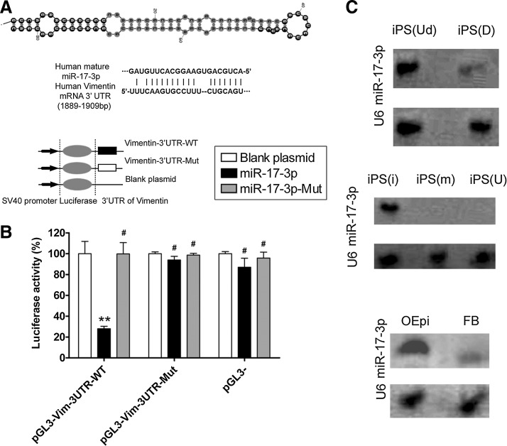
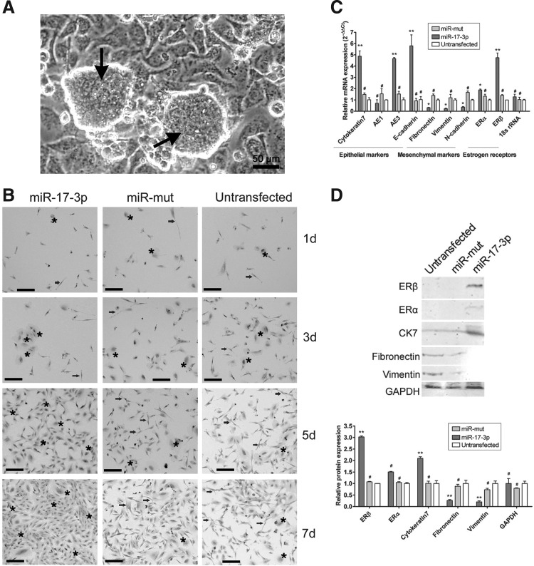
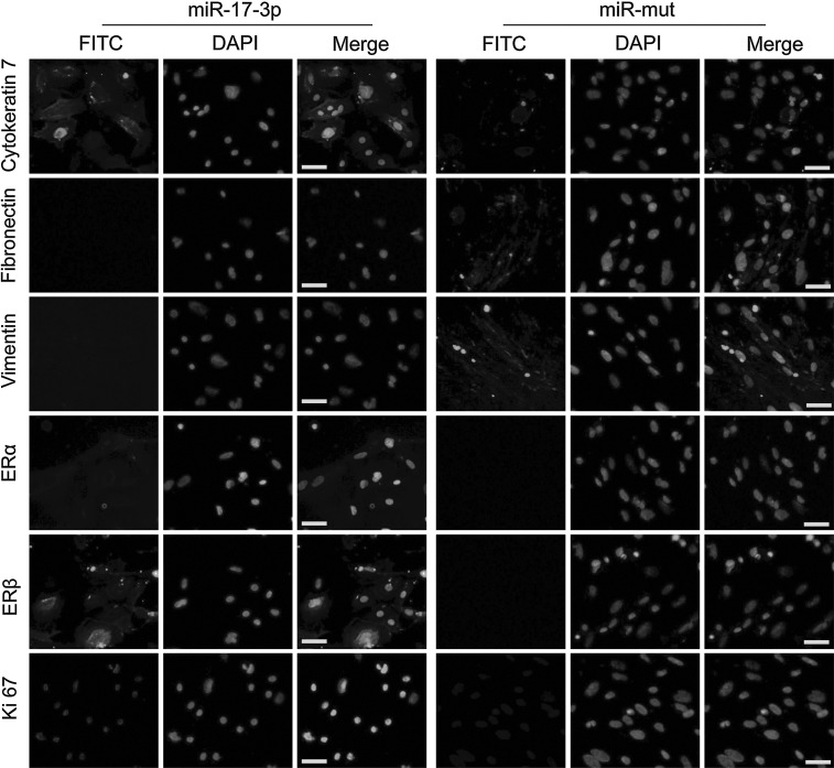
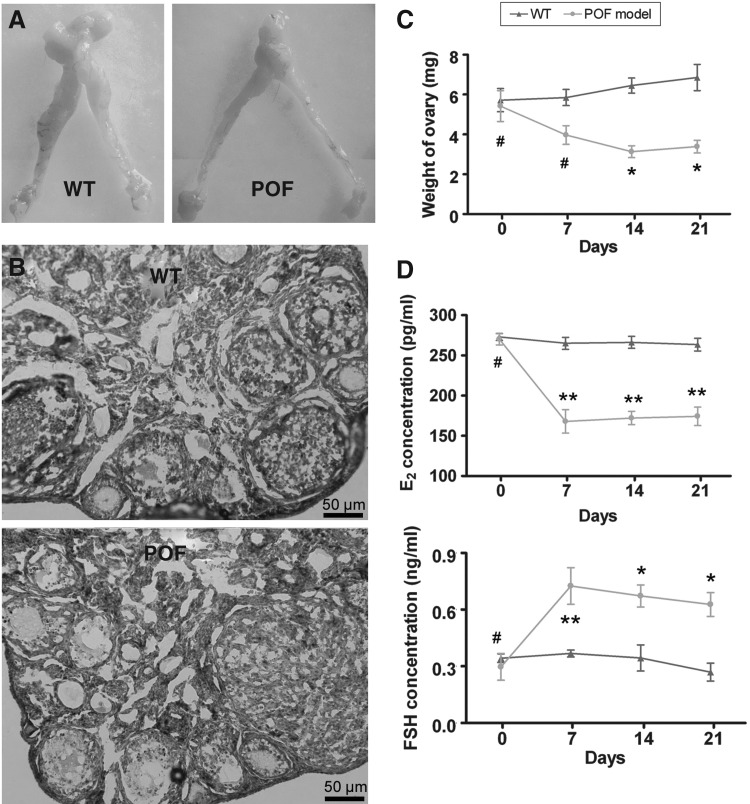
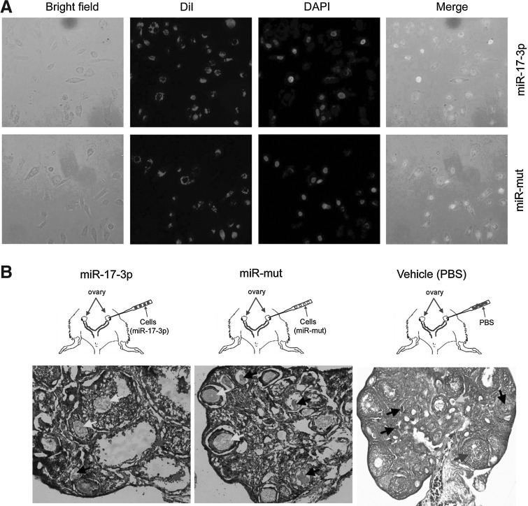
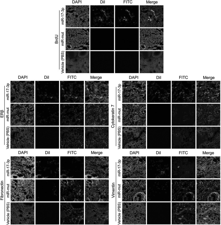
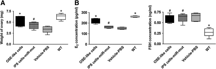
The incidence of premature ovarian failure (POF), a condition causing amenorrhea and hypergonadotropic hypoestrogenism in women before the age of 40, has been increasing in recent years. As an irreversible pathological change, improved treatment strategies for this disease are urgently needed. In this study, a type of microRNA (miR-17-3p) was used to guide the differentiation of human-induced pluripotent stem (iPS) cells into hormone-sensitive ovarian epithelial (OSE)-like cells in vitro. To prevent their morphological transformation into fibroblast-like cells, MiR-17-3p, a microRNA that suppresses vimentin expression, was transfected into human iPS cells. Subsequently, these cells were successfully induced into OSE-like cells in vitro after treatment with estrogen and cell growth factors. Compared with controls, iPS cells transfected with miR-17-3p expressed higher levels of epithelial markers (cytokeratin 7, AE1, AE3, and E-cadherin) and estrogen receptors (ERα and ERβ) while levels of mesenchymal markers (fibronectin, vimentin, and N-cadherin) lowered after the induction. The human iPS cell-derived OSE-like cells were then injected into cyclophosphamide-induced POF model mice to determine their potential benefit as grafts to repair ovarian tissues. The OSE-like cells survived within POF mouse ovaries for at least 14 days in vivo. Compared with the negative controls, expressions of cytokeratin 7 and ERβ proteins were elevated while fibronectin and vimentin levels in ovarian tissues were downregulated in the OSE-like cell transplantation group. Moreover, the ovarian weight and plasma E2 level increased over time in the transplantation with OSE-like cells, compared with control groups. Hence, we can draw the conclusion that iPS cells can be induced to differentiate into OSE-like cells in vitro.


Figures

FIG. 1.
FIG. 1.
Secondary structure of miR-17-3p and its expression in different cells (A) Typical secondary structure of hsa-miR-17-3p precursor miRNAs (pre-miRNAs), contains stem-loop and hairpin structures, with a binding site located in an unstable region with a multi-branching loop-like RNA structure. Complementarity between miR-17-3p and the targeted human vimentin 3′-UTR site (1889–1909 bp downstream), and conserved bases of the mature miR-17-3p target sequence were present in the human vimentin 3′-UTR. (B) Expression of miR-17-3p and its effect on expression of the target gene vimentin was assessed by a luciferase assay. Wild-type (WT) reporter or mutated control luciferase plasmids were transfected into NIH-3T3 cells with miR-17-3p or mut-miR-17-3p expressing lentiviruses. Luciferase activity within the vimentin 3′-UTR sites was inhibited by miR-17-3p (**p<0.01 vs. empty plasmid; #p>0.05 vs. empty plasmid; n=3). (C) Northern blotting results showing expression of miR-17-3p in different cells. In induced pluripotent stem (iPS) cells, a stronger miR-17-3p hybridization signal was observed in human iPS cells at the differentiated stage (D), compared with that at the undifferentiated stage (Ud). In transfected iPS cells, a stronger miR-17-3p hybridization signal was observed in human iPS cells transfected with the miR-17-3p expression plasmid (i), compared with those that were transfected with the mutant miR-17-3p (m) expression plasmid or were untransfected (U). In differentiated iPS cells, a stronger miR-17-3p hybridization signal was seen in ovarian epithelial-like cells derived from iPS cells (OEpi), compared with that in fibroblast cells (FB). U6 RNA was used as a loading control in northern blot assays.
FIG. 2.
FIG. 2.
Expression of multiple cell markers on differentiated cells derived from human iPS cells. (A) Human iPS cells (black arrows) were cultured on HuAECs feeder layers. Scale bar=50 μm. Original magnification, 200×. (B) Morphological evaluation of cell groups (miR-17-3p-transfected iPS cells, miR-mut-transfected iPS cells, and untransfected cells) by H&E staining. Each cell group was induced to differentiate into somatic cells by specific hormones and cell growth factors over 7 days in vitro. The epithelioid-like cells (black stars) gradually increased in number as the in vitro induction time increased, compared with single in the miR-17-3p-transfected iPS cell group. However, in the miR-mut-transfected group and untransfected group, abundant fibroblast-like cells (black arrowheads) were present with fewer interspersed epithelioid-like cells. Scale bar=50 μm. Original magnification, 200×. (C) Quantitative real-time polymerase chain reaction analysis of expression levels of epithelial markers (cytokeratin 7, AE1, AE3, and E-cadherin), mesenchymal markers (fibronectin, vimentin, and N-cadherin), and estrogen receptors (ERα and ERβ) after iPS cells were induced. The miR-17-3p-transfected group expressed epithelial markers and ERβ. Relative mRNA expression is shown after normalization to 18S rRNA, serving as an internal control (**p<0.01 vs. Untransfected; *p<0.05 vs. Untransfected; #p>0.05 vs. Untransfected; n=3). (D) Western blotting showed cytokeratin 7 and ERβ protein expression levels in the miR-17-3p-transfected iPS group were significantly higher than those of the untransfected group. However, fibronectin and vimentin expression levels in the miR-17-3p-transfected iPS group were lower than those of the untransfected group (**p<0.01 vs. Untransfected; #p>0.05 vs. Untransfected; n=3).
FIG. 3.
FIG. 3.
IF staining of several ovarian epithelial cell markers on the 7th day after induction of iPS cells. Expression levels of cytokeratin 7, ERβ, and Ki-67 proteins were elevated in the miR-17-3p-transfected iPS group compared with the miR-mut-transfected iPS group. However, fibronectin and vimentin protein levels were decreased in the miR-17-3p-transfected iPS group compared with the miR-mut-transfected iPS group after differentiation. Scale bar=50 μm. Original magnification, 200×. IF, immunofluorescence staining.
FIG. 4.
FIG. 4.
Establishment of a premature ovarian failure (POF) mouse model and pathological analysis. (A) Female mouse reproductive system. (A) Fallopian tubes were narrowed and the ovaries were atrophied in POF mice model. (B) Pathological observation of ovaries from the WT group and POF group at 2 weeks after injection with cyclophosphamide. The atrophied ovaries of the POF model mice mostly consisted of interstitial cells in a fibrous matrix, with a reduced number of follicles at each stage and an increased number of collapsed oocytes. Scale bar=50 μm. Original magnification, 200×. (C) The weight of ovaries in the POF model mice was found significantly reduced compared with that in normal healthy mice. *p<0.05 versus WT; #p>0.05 versus WT. (D) Plasma E2 levels and follicle stimulating hormone (FSH) levels determined by ELISA at various time points after the injection of cyclophosphamide. **p<0.01 versus WT; #p>0.05 versus WT.
FIG. 5.
FIG. 5.
Fluorescent dye of labeled cells transplanted into ovaries of POF model mice. (A) Each group of cells was labeled with the white fluorescent dye Dil in vitro. (B) H&E staining for pathological analysis of ovaries in each transplant group in POF model mice at 2 weeks after injection of iPS cells or PBS. Follicular atresia and ovarian fibrosis were significantly improved in the miR-17-3p-transfected iPS cells group but not in the miR-mut-transfected iPS cell transplantation group and vehicle group (PBS injected). The black arrows indicate the atretic follicles; white arrows indicate the mature follicles; gray arrows indicate the ovarian granulosa cells. Original magnification, 200×.
FIG. 6.
FIG. 6.
Detection of specific biomarkers and hormone levels and ovarian weight change after cell transplantation. Survival of OSE-like cells in the ovary of a POF mouse model and changes in expression of specific biomarkers IF analysis demonstrated that Dil, a marker of transplanted cells, was expressed at higher levels in graft OSE-like cells in the mouse ovaries. The transplanted cells survived for at least 2 weeks in vivo. Moreover, BrdU incorporation and IF staining indicated that transplanted OSE-like cells had undergone normal cell division. However, in both the miR-mut-transfected iPS cell transplantation group and the vehicle group (PBS injected), Dil (white fluorescent, RF) and BrdU (white fluorescent) signals were not found. Thus, only OSE-like cells could be detected along the injection tract in POF mouse ovaries. Original magnification, 200×. In addition, cytokeratin 7 and ERβ protein levels were elevated in ovarian tissues of the group transplanted with RF-positive OSE-like cells compared with those transplanted with miR-mut iPS cells or vehicle group (PBS injected). However, fibronectin and vimentin protein levels were decreased in ovarian tissues of the group transplanted with RF-positive OSE-like cells compared with the other two groups. Original magnification, 200×.
FIG. 7.
FIG. 7.
Detection of hormone levels and ovarian weight after cell transplantation. (A) Ovarian weight in POF model mice transplanted with OSE-like cells increased significantly compared with that in the other two groups 14 days after transplantation (*p<0.05 vs. vehicle group; #p>0.05 vs. vehicle group; n=8). (B) Plasma E2 levels increased over time in the POF model mice transplanted with OSE-like cells compared with the other two groups. However, no significant difference in plasma FSH concentration was observed among the three groups (#p> 0.05 vs. vehicle group; n=8).

References

    1. Auersperg N. Wong A.S. Choi K.C. Kang S.K. Leung P.C. Ovarian surface epithelium: biology, endocrinology, and pathology. Endocr Rev. 2001;22:255–288. - PubMed
    1. Bandyopadhyay S. Chakrabarti J. Banerjee S. Pal A.K. Goswami S.K. Chakravarty B.N., et al. Galactose toxicity in the rat as a model for premature ovarian failure: an experimental approach readdressed. Hum Reprod. 2003;18:2031–2038. - PubMed
    1. Cheng C.W. Wang H.W. Chang C.W. Chu H.W. Chen C.Y. Yu J.C., et al. MicroRNA-30a inhibits cell migration and invasion by downregulating vimentin expression and is a potential prognostic marker in breast cancer. Breast Cancer Res Treat. 2012a;134:1081–1093. - PubMed
    1. Cheng W. Liu T. Jiang F. Liu C. Zhao X. Gao Y., et al. microRNA-155 regulates angiotensin II type 1 receptor expression in umbilical vein endothelial cells from severely pre-eclamptic pregnant women. Int J Mol Med. 2011;27:393–399. - PubMed
    1. Cheng W. Liu T. Wan X. Gao Y. Wang H. MicroRNA-199a targets CD44 to suppress the tumorigenicity and multidrug resistance of ovarian cancer-initiating cells. FEBS J. 2012b;279:2047–2059. - PubMed

Publication types

Substances