. 2013 Nov;84(5):453-63.
doi: 10.1111/cge.12257.

A systematic approach to assessing the clinical significance of genetic variants

Affiliations

A systematic approach to assessing the clinical significance of genetic variants

H Duzkale et al. Clin Genet. 2013 Nov.

Abstract

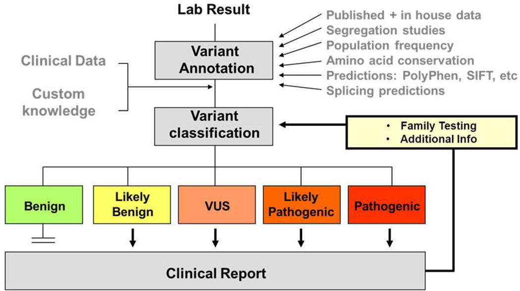
Molecular genetic testing informs diagnosis, prognosis, and risk assessment for patients and their family members. Recent advances in low-cost, high-throughput DNA sequencing and computing technologies have enabled the rapid expansion of genetic test content, resulting in dramatically increased numbers of DNA variants identified per test. To address this challenge, our laboratory has developed a systematic approach to thorough and efficient assessments of variants for pathogenicity determination. We first search for existing data in publications and databases including internal, collaborative and public resources. We then perform full evidence-based assessments through statistical analyses of observations in the general population and disease cohorts, evaluation of experimental data from in vivo or in vitro studies, and computational predictions of potential impacts of each variant. Finally, we weigh all evidence to reach an overall conclusion on the potential for each variant to be disease causing. In this report, we highlight the principles of variant assessment, address the caveats and pitfalls, and provide examples to illustrate the process. By sharing our experience and providing a framework for variant assessment, including access to a freely available customizable tool, we hope to help move towards standardized and consistent approaches to variant assessment.

Keywords: (4−9) clinical interpretation; gain-of-function (GOF); genetic variant; loss of function (LOF); next-generation sequencing (NGS); sequence analysis; variant assessment; variant of uncertain significance (VUS).


Conflict of interest statement

Conflict of interest statement

HD, JS, HM, MAK, TJP, BHF, HLR and MSL are employed by fee-for-service laboratories performing clinical sequencing services. Several individuals serve on advisory boards or in other capacities for companies providing sequencing or other genetic services (HLR – BioBase, Clinical Future, Complete Genomics, GenomeQuest, Illumina, Ingenuity, Knome, Omicia; BF – InVitae; JS – LabCorp).

Figures

Figure 1
Figure 1
Variant assessment workflow. Genetic variants identified by laboratory testing are annotated with information from various sources including publications, computational prediction algorithms, and public, collaborative and internal databases. After evaluation of all pertinent information in conjunction with patient specific clinical and family information, a professionally trained individual will classify the variant into one of the five clinical categories and combine all variants for a clinical report.
Figure 2
Figure 2
A. Reference transcript selection. Two transcripts for OTOF are shown: NM_194248, the longest transcript selected as the primary transcript, and NM_194322, a shorter alternate transcript. Position g.26799794 (grey box) is non-coding in NM_194248 (c.2250–80) but coding in NM_194322 (c.66); therefore NM_194322 should be selected while evaluating this variant. Figure 2B. Phasing multiple variants. Two variants are present at positions c.2401 and c.2402 in OTOF (NM_194248). The top traces show the chromatographs from Sanger sequencing with the consensus reference sequence shown underneath. Representative aligned NGS reads are shown below. Grey bars represent reference sequence with variants highlighted in red. The bottom schematic shows associated OTOF coding exons (rectangles) and the reference amino acid sequence. The arrow indicates the 5’ to 3’ direction. The c.2401G>T (p.Glu801*) is listed in dbSNP (rs75624587) and ESP as a nonsense variant but with an allele frequency of 10% in African Americans. However, NGS reads reveal that the variants are in cis and should therefore be named c.2401_2402delinsTT (p.Glu891Leu). Figure 2C. Segregation analysis with incomplete penetrance. A family with hypertrophic cardiomyopathy is shown. Affected individuals are indicated by filled squares (males) or circles (females). Mutation-positive individuals are indicated by a “+”, while mutation-negative individuals are indicated by a “−“. All mutation-positive individuals are affected, with the exception of individual II-1. Because HCM can display reduced penetrance, individual II-1 would not be considered a non-segregation. Figure 2D. Conservation based on multiple species alignment. An example of alignment of TNNC1 in UCSC Genome Browser is shown. Tree shrew sequence shows poor alignment (red box). Arrows point to non-conserved residues. Figure 2E. Conflicting computational predictions of a missense variant. The results of multiple computational tools are captured in the VAT. They provide conflicting predictions for the NM_000366:c.688G>A (p.Asp230Asn) variant in TPM1, suggesting that at least some tools are not reliable. Figure 2F. Predicted splicing effect of a coding variant. Splicing prediction tools indicate that the NM_022124:c.5712G>A variant in CDH23, which affects the last base in exon 43, may impact splicing. However, not all programs agree in the potential effect on splicing, and they cannot predict whether it would lead to exon skipping, intron retention or use of cryptic splice sites.
Figure 2
Figure 2
A. Reference transcript selection. Two transcripts for OTOF are shown: NM_194248, the longest transcript selected as the primary transcript, and NM_194322, a shorter alternate transcript. Position g.26799794 (grey box) is non-coding in NM_194248 (c.2250–80) but coding in NM_194322 (c.66); therefore NM_194322 should be selected while evaluating this variant. Figure 2B. Phasing multiple variants. Two variants are present at positions c.2401 and c.2402 in OTOF (NM_194248). The top traces show the chromatographs from Sanger sequencing with the consensus reference sequence shown underneath. Representative aligned NGS reads are shown below. Grey bars represent reference sequence with variants highlighted in red. The bottom schematic shows associated OTOF coding exons (rectangles) and the reference amino acid sequence. The arrow indicates the 5’ to 3’ direction. The c.2401G>T (p.Glu801*) is listed in dbSNP (rs75624587) and ESP as a nonsense variant but with an allele frequency of 10% in African Americans. However, NGS reads reveal that the variants are in cis and should therefore be named c.2401_2402delinsTT (p.Glu891Leu). Figure 2C. Segregation analysis with incomplete penetrance. A family with hypertrophic cardiomyopathy is shown. Affected individuals are indicated by filled squares (males) or circles (females). Mutation-positive individuals are indicated by a “+”, while mutation-negative individuals are indicated by a “−“. All mutation-positive individuals are affected, with the exception of individual II-1. Because HCM can display reduced penetrance, individual II-1 would not be considered a non-segregation. Figure 2D. Conservation based on multiple species alignment. An example of alignment of TNNC1 in UCSC Genome Browser is shown. Tree shrew sequence shows poor alignment (red box). Arrows point to non-conserved residues. Figure 2E. Conflicting computational predictions of a missense variant. The results of multiple computational tools are captured in the VAT. They provide conflicting predictions for the NM_000366:c.688G>A (p.Asp230Asn) variant in TPM1, suggesting that at least some tools are not reliable. Figure 2F. Predicted splicing effect of a coding variant. Splicing prediction tools indicate that the NM_022124:c.5712G>A variant in CDH23, which affects the last base in exon 43, may impact splicing. However, not all programs agree in the potential effect on splicing, and they cannot predict whether it would lead to exon skipping, intron retention or use of cryptic splice sites.

References

    1. Richards CS, Bradley LA, Amos J, Allitto B, Grody WW, Maddalena A, McGinnis MJ, Prior TW, Popovich BW, Watson MS, et al. Standards and guidelines for CFTR mutation testing. Genet Med. 2002;4:379–391. - PubMed
    1. Huang T. Next generation sequencing to characterize mitochondrial genomic DNA heteroplasmy. Curr Protoc Hum Genet. 2011;Chapter 19(Unit19):Unit18. - PMC - PubMed
    1. Valencia CA, Ankala A, Rhodenizer D, Bhide S, Littlejohn MR, Keong LM, Rutkowski A, Sparks S, Bonnemann C, Hegde M. Comprehensive mutation analysis for congenital muscular dystrophy: a clinical PCR-based enrichment and next-generation sequencing panel. PLoS One. 2013;8:e53083. - PMC - PubMed
    1. Richards CS, Bale S, Bellissimo DB, Das S, Grody WW, Hegde MR, Lyon E, Ward BE. ACMG recommendations for standards for interpretation and reporting of sequence variations: Revisions 2007. Genet Med. 2008;10:294–300. - PubMed
    1. Kearney HM, Thorland EC, Brown KK, Quintero-Rivera F, South ST Working Group of the American College of Medical Genetics Laboratory Quality Assurance, C. American College of Medical Genetics standards and guidelines for interpretation and reporting of postnatal constitutional copy number variants. Genet Med. 2011;13:680–685. - PubMed

Publication types